Positioning and partition method for human tissue cell two-photon microscopic image

A microscopic image and human tissue technology, applied in the field of image processing, can solve problems such as unsatisfactory segmentation results, segmentation results that cannot be used for image classification, pattern recognition, etc., to increase efficiency, reduce evolution time, and increase accuracy.

Active Publication Date: 2013-10-09
FUJIAN NORMAL UNIV
View PDF2 Cites 18 Cited by
  • Summary
  • Abstract
  • Description
  • Claims
  • Application Information

AI Technical Summary

Problems solved by technology

Due to the fact that there are very few studies on two-photon microscopic images of cells at home and abroad, and the existing methods for segmenting two-photon microscopic images of human tissue cells are not ideal, the segmentation results cannot be used for subsequent image classification and model analysis. Identification and other processing

Method used


Image

  • Positioning and partition method for human tissue cell two-photon microscopic image
  • Positioning and partition method for human tissue cell two-photon microscopic image
  • Positioning and partition method for human tissue cell two-photon microscopic image

Examples


Embodiment 1

[0056] Localization and segmentation of two-photon microscopic images of human nasopharyngeal epithelial cells

[0057] The specific process is as follows:

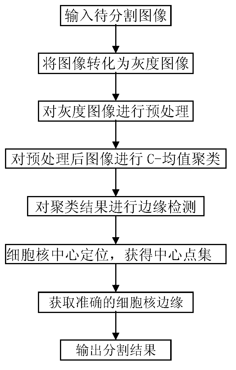
[0058] Step 1. Input the two-photon microscopy image to be segmented and convert the image into a grayscale image

[0059] will be obtained using two-photon microscopy as Figure 5 (a) The original image of human nasopharyngeal epithelial cells showing green color is converted into a grayscale image, and the converted grayscale image is as follows Figure 5 (b) shown.

[0060] Step 2. Preprocess the grayscale image to reduce noise and increase contrast

[0061] Depend on Figure 5 (b) It can be seen that the noise of the original grayscale image is relatively serious, and the interference objects are mostly granular. At the same time, the contrast in some areas is low, and it is difficult to distinguish the cytoplasm or nucleus with the naked eye. Therefore, proper preprocessing of the original grayscale image is re...


PUM


Abstract

The invention discloses a positioning and partition method for a human tissue cell two-photon microscopic image, and belongs to the technical field of image processing. The positioning and partition method for the human tissue cell two-photon microscopic image mainly solves the problem that too many errors occur when the human tissue cell two-photon microscopic images are partitioned by means of the prior art. The positioning and partition method comprises the steps of preprocessing, namely converting an image to be partitioned into a grey-scale image, clustering the preprocessed images and obtaining an edge graph of the image, conducting centralized positioning with the edge graph, positioning the centers of cell nucleuses accurately, obtaining a point set of the cell nucleus centers, and finally obtaining the edges of the cell nucleuses accurately combining an active contour model. Compared with the prior art, the positioning and partition method for the human tissue cell two-photon microscopic image has the advantages of being accurate in edge extraction, high in positioning efficiency, short in evolution time and the like. Further, the disturbance of granular noises and uniform brightness distribution in the image is avoided, and the positioning and partition method can be used for extracting the edges of the cell nucleuses in the cell two-photon microscopic image.

Description

technical field [0001] The invention belongs to the technical field of image processing, and relates to a method for positioning and segmenting two-photon microscopic images of human tissue cells. Background technique [0002] Image segmentation is an important topic in the field of image processing. Effective and reasonable image segmentation can provide favorable conditions for subsequent processing such as image classification and pattern recognition. For a long time, scholars at home and abroad have conducted a lot of research on the problem of medical cell image segmentation, and have proposed thousands of image segmentation algorithms. However, due to the complexity of practical problems, a segmentation algorithm suitable for all kinds of images has not been proposed so far. [0003] The traditional segmentation methods of cell microscopic images can be divided into threshold segmentation, edge detection and region segmentation methods. These methods have good segment...

Claims


Application Information

Patent Timeline
Patent Type & Authority Applications(China)
IPC IPC(8): G06T7/00G06T5/00
Inventor 陈冠楠胡恒阳陈荣黄祖芳刘垚蔡坚勇王平林居强冯尚源
Owner FUJIAN NORMAL UNIV
Why Eureka
Try Eureka