Method for acquisition of subtraction angiograms

A technology before image and radiography, applied in image enhancement, image analysis, image data processing, etc., can solve the problems of spatial resolution reduction, negative impact of motion compensation accuracy, error accumulation, etc., and achieve the effect of improving diagnostic value

Inactive Publication Date: 2014-01-29
ZAKRYTOE AKCIONERNOE OBSHCHESTVO IMPULS
View PDF5 Cites 8 Cited by
  • Summary
  • Abstract
  • Description
  • Claims
  • Application Information

AI Technical Summary

Problems solved by technology

Multiple spatial frequency analyzes of images encountered in clinical practice show that many images contain essential details that only result in a loss of spatial resolution
In this case, the above method can give accurate results for the determined movement, but after the determined movement, the error will be accumulated
[0039] Furthermore, according to known methods, the function of the coordinate transformation is smooth in the ROI, which has a negative impact on the accuracy of motion compensation if there are at least two moving parts in the ROI with clear edges

Method used


Image

  • Method for acquisition of subtraction angiograms
  • Method for acquisition of subtraction angiograms
  • Method for acquisition of subtraction angiograms

Examples


Embodiment

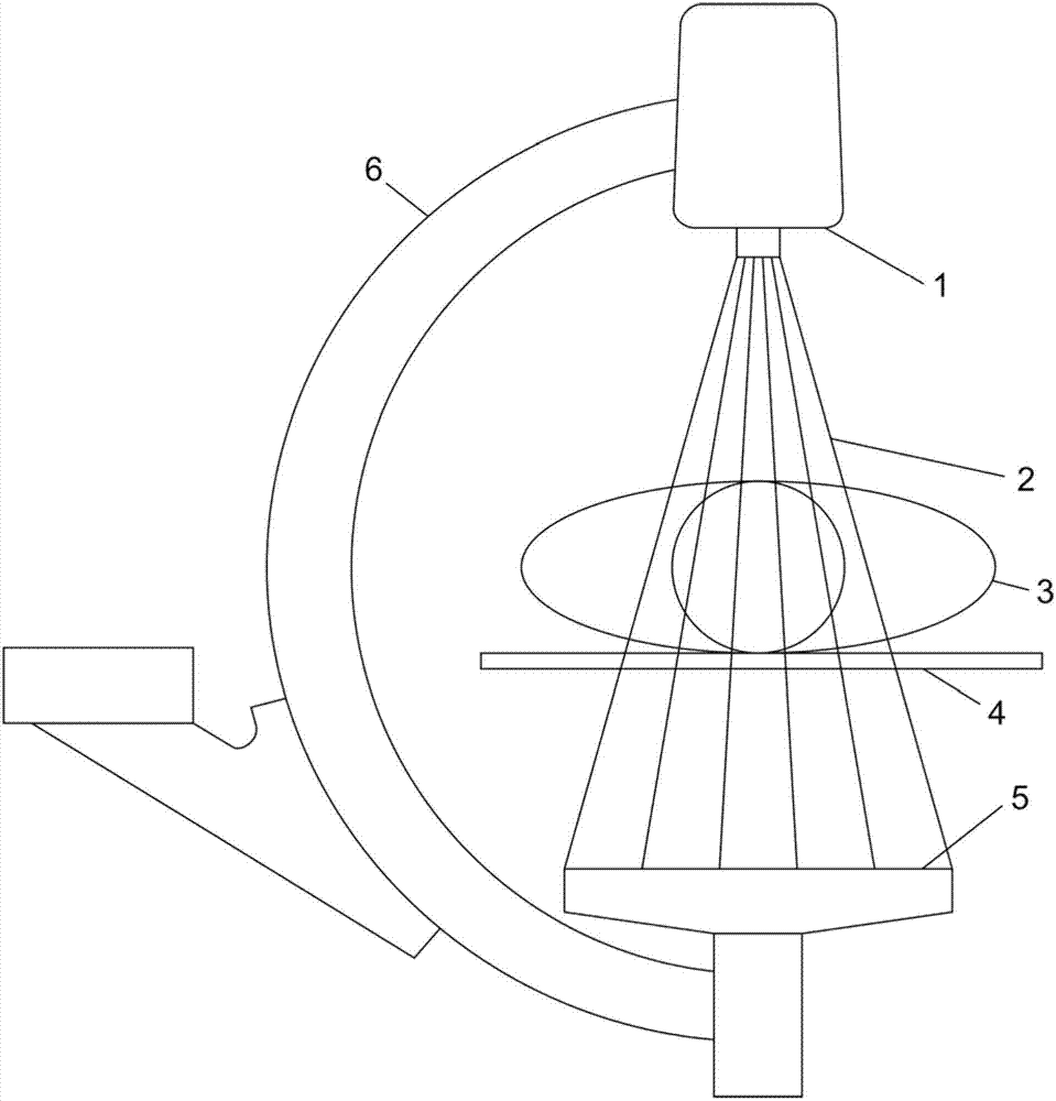
[0081] The x-ray images are obtained by using an x-ray system, the fixed parts of which are not projected onto the working area of ​​the detector. X-ray irradiation is performed to obtain 2 to 3 digital images of the pre-contrast sequence. In the first digital image of the pre-contrast image sequence, feature details are searched. To do this, proceed as follows:

[0082] 1. For images, a multiscale representation consisting of 4 layers is formed. The first layer is the image itself. Then, to form each next layer, the image is subsampled by a factor of 2 using a digital smoothing filter with a window size of 3×3.

[0083] 2. At each layer of the multi-scale image, feature details are selected. To do this, first calculate the first partial derivatives of the brightness in the horizontal and vertical directions:

[0084] I x ′ = ∂ I ∂ ...


PUM


Abstract

The invention relates to the field of digital processing of X-ray images and can be used in digital subtraction angiography to compensate for impact of involuntary patient movement and movement of internal organs on image of the vascular system. The technical result of the claimed invention is the improvement of diagnostic value of subtraction angiographic images by eliminating artifacts caused by the motility of anatomical structures. Technical result is achieved by the that at the stage of digital images registration a search for characteristic details is performed for each image. According to the shift of the said details determine expectable shift of a patient organs. Then perform segmentation of image from the pre-contrast series in the region of homogenous warping ; for each region a geometrical transformation is calculated and corresponding geometrical transformations are performed for each region of a series of pre-contrast digital images. This means vessels visibility enhancement and treatment and diagnostic improvement.

Description

technical field [0001] The invention relates to the field of digital X-ray image processing, and can be applied to digital subtraction angiography to compensate the influence of involuntary movement of patients and normal movement of internal organs on images of blood vessels. Background technique [0002] Angiography is a medical imaging technique that visualizes the inside of blood vessels in a patient by injecting a contrast agent into them. In an angiogram, in addition to blood vessels, images of surrounding organs and tissues can be seen, which often interfere with the identification of blood vessels filled with contrast agent. To reduce the visibility of surrounding tissue during digital subtraction angiography (DSA), a pre-contrast image is generated, which is a region of interest in a patient acquired before contrast agent is injected into the vasculature. , ROI) X-ray image. A sequence of images showing the path of the injected contrast agent through the vessel of...

Claims


Application Information

Patent Timeline
Patent Type & Authority Applications(China)
IPC IPC(8): G06T5/50
CPCG06T2207/20012G06T2207/10116G06T5/50G06T2207/20016G06T2207/20224G06T7/0026G06T7/00G06T2207/30101G06T7/32A61B6/00G06T5/00
Inventor A·S·阿法纳先科
Owner ZAKRYTOE AKCIONERNOE OBSHCHESTVO IMPULS
Why Eureka
Try Eureka