Medical Image Processing Method Based on Network Phase Synchronization

A medical imaging and phase synchronization technology, applied in the field of image processing, can solve the problems of blurred medical images, large individual differences, unevenness, etc., and achieve the effect of small calculation amount, fast calculation speed, and improved diagnosis and recognition rate.

Active Publication Date: 2016-07-06
XIDIAN UNIV
View PDF3 Cites 0 Cited by
  • Summary
  • Abstract
  • Description
  • Claims
  • Application Information

AI Technical Summary

Problems solved by technology

[0004] The purpose of the present invention is to propose a medical image processing method based on network phase synchronization to solve the problem of over-segmentation in existing methods and improve the diagnostic recognition rate of medical images for the problems of blurred, uneven, and large individual differences in medical images

Method used


Image

  • Medical Image Processing Method Based on Network Phase Synchronization
  • Medical Image Processing Method Based on Network Phase Synchronization
  • Medical Image Processing Method Based on Network Phase Synchronization

Examples


Embodiment Construction

[0024] refer to figure 1 , the present invention is based on the medical image processing method of network phase synchronization, comprising the following steps:

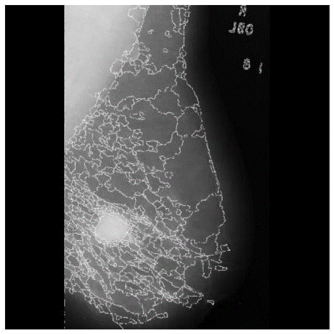
[0025] Step 1, extract the features of the original medical image pixels and perform watershed segmentation on the features of the pixels.

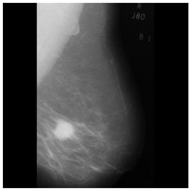
[0026] (1a) Input the original medical image, this example selects as figure 2 The original mammogram image shown, the size is 1024×1024;

[0027] (1b) Extract the features based on the gray level co-occurrence matrix. For any pixel point i, extract the 12-dimensional features of contrast, consistency, and energy in the four directions of [0, 45, 90, 135], and then extract the features based on non-sampling wavelet transform 10-dimensional feature, the above-mentioned 12-dimensional feature and 10-dimensional feature are combined to form a 22-dimensional vector, which is used as the feature of the i-th pixel;

[0028] (1c) Repeat step (1b) for all pixels in the image to ob...


PUM


Abstract

The invention discloses a medical image processing method based on network phase synchronization, which mainly solves the problems of low diagnostic recognition rate and poor processing of local details in the existing medical image processing method. The implementation steps are: (1) read in the original medical image, extract the features of the pixels, and perform watershed segmentation; (2) calculate the similarity between the segmented regions; (3) use the region as a node, and the similarity as the network weight (4) Randomly generate the initial phase of the network, iterate the phase until it is stable, and extract the stable node phase; (5) Use the stable phase to classify the nodes first, and then calculate the node slope, according to the slope Classify the nodes; (6) Redistribute the misclassified nodes and mark the category numbers on the pixels; (7) Assign different gray values ​​to different types of pixels to obtain the segmented image. Compared with the existing method, the invention improves the diagnostic recognition rate of the medical image.

Description

technical field [0001] The invention belongs to the field of image processing, relates to the processing of medical images, and can be used for monitoring disease distribution, researching pathogenesis and assisting disease diagnosis. Background technique [0002] Medical images include CT, positron emission tomography PET, single photon radiation tomography SPECT, nuclear magnetic resonance imaging MR, and images obtained by other medical imaging equipment. Due to the complexity and diversity of medical images, as well as the differences in imaging principles and tissue characteristics of medical imaging, and the formation of images is affected by noise, field offset effects, local body effects, and tissue motion, medical imaging and general Compared with the image, it has the characteristics of blur and inhomogeneity. In addition, the structure and shape of human anatomy are complex, and the differences between different individuals are large, which also makes the problem...

Claims


Application Information

Patent Timeline
Patent Type & Authority Patents(China)
IPC IPC(8): G06T7/00
Inventor 吴建设马文萍焦杨冯婕马晶晶王爽侯彪公茂果
Owner XIDIAN UNIV
Why Eureka
Try Eureka