System and method for capturing and visualizing mandibular three-dimensional motion

A three-dimensional movement and mandibular technology, applied in the field of medical inspection, can solve the problems of inconvenient operation, high price, and inability to quantitatively analyze the movement of the condyle.

Active Publication Date: 2014-04-02
SUN YAT SEN UNIV
View PDF8 Cites 24 Cited by
  • Summary
  • Abstract
  • Description
  • Claims
  • Application Information

AI Technical Summary

Problems solved by technology

However, the overlap and projection angle of X-ray images cannot quantitatively analyze the movement of certain points on the condyle
In 1921, B. McCollum developed a mechanical mandibular movement recorder, which recorded the movement of the mandible through the splint, facebow and tracing pen board. After many improvements, it has become the main research method of current clinical research, but it is still too cumbersome. and complex, not conducive to promotion
In the 1980s, the American Grbh company successfully developed the condyle motion tracer, which combined the classic condyle motion trace with the computer, but it still has its limitations, because the oute

Method used


Image

  • System and method for capturing and visualizing mandibular three-dimensional motion
  • System and method for capturing and visualizing mandibular three-dimensional motion
  • System and method for capturing and visualizing mandibular three-dimensional motion

Examples


Embodiment 1

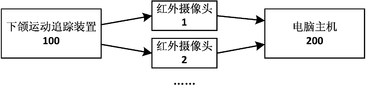
[0040] see figure 1 , is a schematic structural diagram of the mandibular three-dimensional motion capture and visualization system provided in Embodiment 1 of the present invention.

[0041] In this embodiment, the mandibular three-dimensional motion capture and visualization system includes: a mandibular motion tracking device 100 designed and printed using a three-dimensional CT model, at least two infrared cameras (such as figure 1 Infrared camera 1 and infrared camera 2) and a host computer 200 for analyzing camera recording data.

[0042] Among them, CT is the abbreviation of Computed Tomography (Computed Tomography). According to the difference in the absorption and transmittance of X-rays by different tissues of the human body, it uses highly sensitive instruments to measure the human body, and then inputs the data obtained from the measurement into Computer, after the computer processes the data, it can take a cross-sectional or three-dimensional image of the inspect...

Embodiment 2

[0055] see Figure 4 , is a flow chart of the steps of the method for capturing and visualizing the three-dimensional motion of the mandible provided in Embodiment 2 of the present invention.

[0056] In this embodiment, a mandibular three-dimensional motion capture and visualization method is realized by using the mandibular three-dimensional motion capture and visualization system provided in the first embodiment. Specifically, such as Figure 4 As shown, the method includes the following steps:

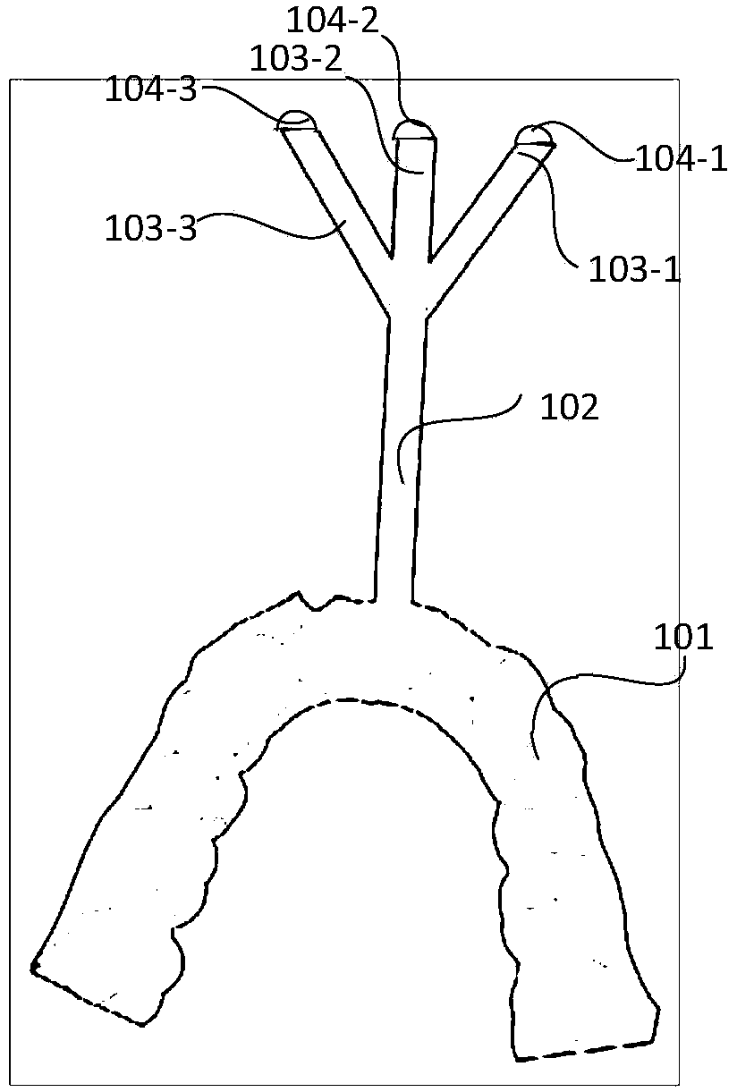
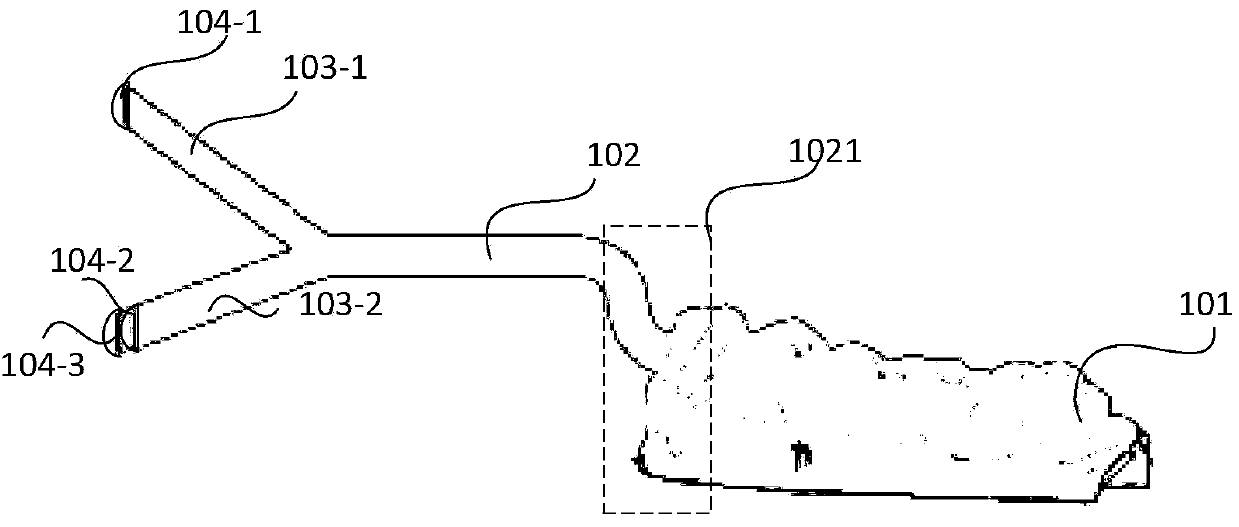
[0057] Step S401: After printing out the mandibular movement tracking device using the 3D CT model, fix the rear-end fixing device on the tester's mandibular incisors, place the three non-coplanar marker bars and The corresponding markers are exposed outside the mouth and move together with the lower jaw; preferably, the rear-end fixing device and the intermediate connecting rod are designed using a three-dimensional CT model according to the shape characteristics of the teeth of...


PUM


Abstract

The invention discloses a system for capturing and visualizing mandibular three-dimensional motion. The system comprises a mandibular motion tracking device for designing and printing through a three-dimensional CT (Computed Tomography) model, at least two infrared cameras, and a host computer for analyzing shooting record data, wherein the mandibular motion tracking device comprises a rear end fixing device, a medium connecting rod, and three marking rods, wherein the three marking rods are arranged at the front end and are not co-planar; one end of each of the marking rods is connected to the medium connecting rod and while the other end of each of the marking rods is provided with a marker; the rear end fixing device is used for designing and printing through the three-dimensional CT model according to the external physical characteristics of lower teeth; the markers expose from the mouth and move together with the mandible; the host computer is used for recovering the overall motion of the mandible of a tested subject so as to determine whether the mandible motion and occlusal contacts are normal. The invention also provides a method for capturing and visualizing the mandibular three-dimensional motion. According to the technical scheme, the system and method have the advantages of low cost, high testing accuracy and the like, and is convenient to operate.

Description

technical field [0001] The invention relates to a medical detection technology, in particular to a mandibular three-dimensional motion capture and visualization system and method. Background technique [0002] Human mandibular movement is a complex three-dimensional movement involving teeth, masticatory muscles and temporomandibular joints. Its movement pattern is closely related to the health of the oral and jaw system. The characteristics of mandibular movement can be used to analyze and understand chewing, swallowing and language. And other functions, for the analysis and diagnosis of temporomandibular joint lesions and judgment of curative effect. Pathophysiological factors such as mandibular incisor trajectory, molar trajectory, bilateral condyle trajectory and the myoelectricity of each masticatory muscle in various movements are the basis for formulating a rehabilitation program for occlusal disease, and they are also the basis for the design of capture motion markers...

Claims


Application Information

Patent Timeline
IPC IPC(8): A61B5/11A61C19/045
Inventor 许跃
Owner SUN YAT SEN UNIV
Why Eureka
Try Eureka