
Method and device for reading medical image files

A technology of medical images and files, applied in the field of computing technology in the medical field, can solve the problems that it is difficult for hospitals and doctors to obtain medical image files, and achieve the effect of improving transmission speed and eliminating synchronization dependence

Inactive Publication Date: 2014-04-09
SIEMENS AG
View PDF4 Cites 7 Cited by
  • Summary
  • Abstract
  • Description
  • Claims
  • Application Information

AI Technical Summary

Problems solved by technology

However, due to the large size of medical image files, hospitals, especially those in economically backward areas, cannot be connected to the Internet at any time, so it is difficult to ensure that hospitals and doctors can obtain the required medical image files in a timely manner.
Therefore, it is difficult for the existing technology to share medical image files between hospitals and doctors in different hospitals.
In addition, there is currently a lack of a network system for trained doctors to access medical imaging files

Method used


Image

  • Method and device for reading medical image files
  • Method and device for reading medical image files
  • Method and device for reading medical image files

Examples


Embodiment Construction

[0017] In order to make the purpose, technical solutions and advantages of the embodiments of the present invention clearer, the following examples are used to further describe the embodiments of the present invention in detail.

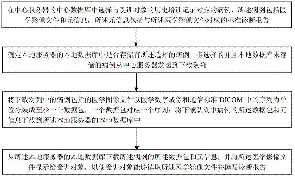
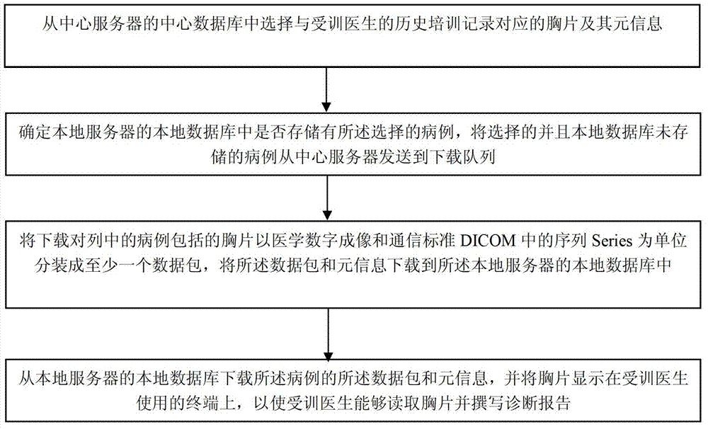
[0018] Such as figure 1 As shown, the method for reading medical image files provided by an embodiment of the present invention includes the following steps.

[0019] Step 11, select a case corresponding to the historical training records of the trainee in the central database of the central server, the case includes a medical image file and meta information, the meta information includes a standard diagnosis report corresponding to the medical image file.

[0020] Here, the standard diagnosis report may be a diagnosis report written by an experienced doctor, for example, a report written for experts from a tertiary hospital. Medical image files include B-ultrasound, CT, etc. In addition to the standard diagnosis report, meta-information can also i...


PUM


Abstract

A method for reading medical image files comprises the following steps of selecting a clinical history, which corresponds to a historical training record of a trained object and comprises a medical image file and meta-information comprising a standard diagnostic report corresponding to the medical image file, from a center database; judging whether the selected clinical history is stored in a local database or not; transmitting the selected clinical history which is not stored in the local database to a downloading queue from the center database; packaging the medicine image file in the clinical history in the downloading queue into at least one data package by using sequences SERIES in a medical digital imaging and communication standard DICOM; enabling the data packages to correspond to the sequences one by one; downloading the data packages of the clinical history in the downloading queue and meta-information to the local database; downloading the data packages and the meta-information of the clinical history from the local database; and displaying the medical image file to the trained object.

Description

technical field [0001] The invention relates to the application of computer technology in the medical field, in particular to a method and device for reading medical image files. Background technique [0002] Medical imaging diagnosis (such as B-ultrasound, CT, X-ray film, etc.) is an important part of clinical diagnosis and plays a pivotal role. The correctness of medical imaging diagnosis is directly related to whether patients can receive timely, reasonable and effective treatment. However, at present, medical image files are rarely shared between hospitals. On the one hand, medical image files are usually very large, and it is difficult to store a large number of them on computers used by doctors or on servers of hospitals. On the other hand, at present, the transmission of medical images is mainly realized by HyperText Transfer Protocol (HyperText Transfer Protocol, HTTP), File Transfer Protocol (File Transfer Protocol, FTP) and so on. As far as the implementation of ...

Claims


Application Information

Patent Timeline
IPC IPC(8): G06F19/00
Inventor 姜恒
Owner SIEMENS AG
Learn More