Wireless three-dimensional ultrasound imaging method and device
A technology of three-dimensional ultrasound and imaging methods, which is applied in ultrasonic/sonic/infrasonic diagnosis, sonic diagnosis, infrasound diagnosis, etc., and can solve the problems of difficulty in obtaining ultrasonic image data, expensive commercial telemedicine systems, and lack of telemedicine. Achieve the effect of strong portability, enhanced portability and mobility, and increased freedom
- Summary
- Abstract
- Description
- Claims
- Application Information
AI Technical Summary
Problems solved by technology
Method used
Image
Examples
Embodiment 1
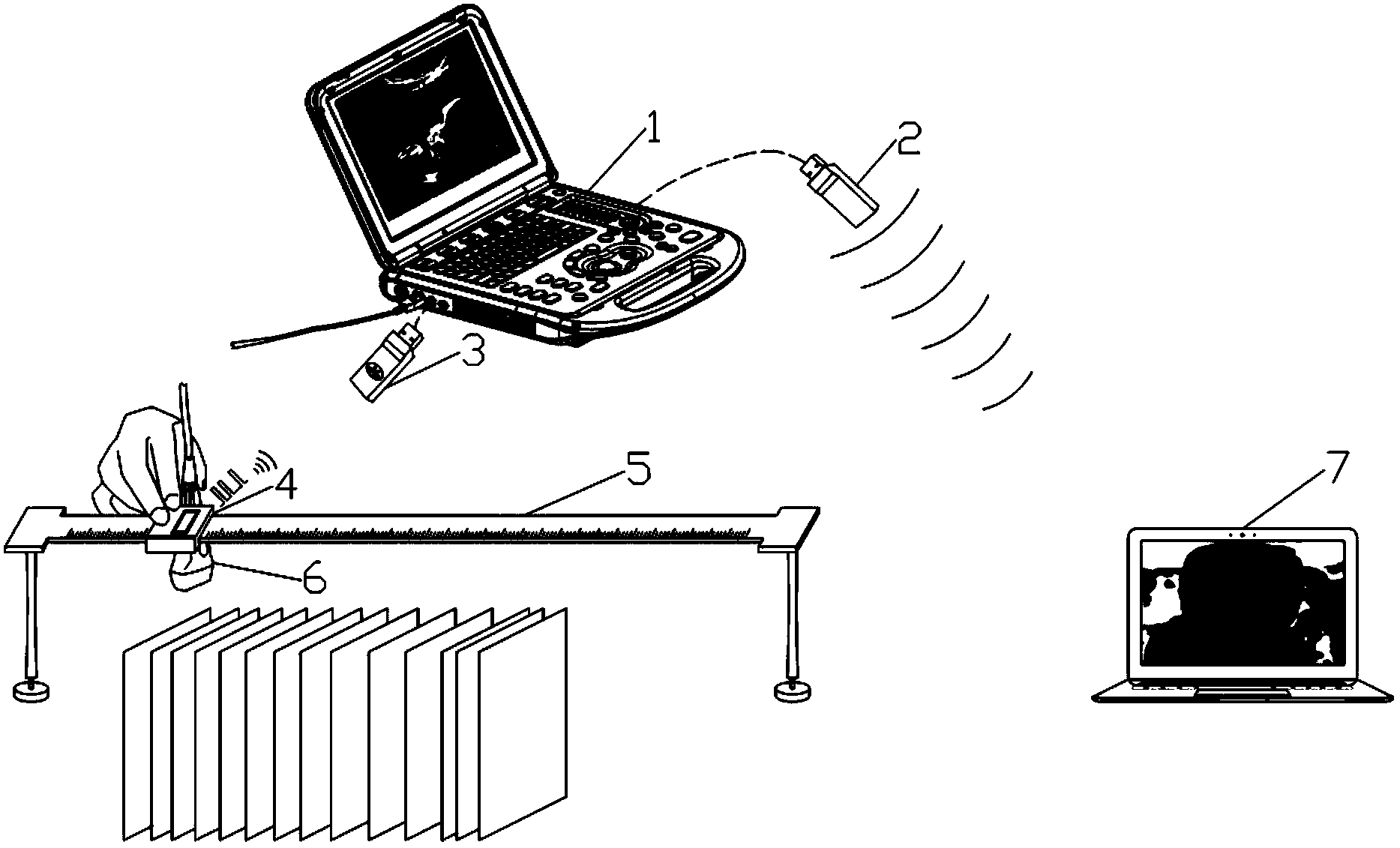
[0036] Such as figure 1 As shown, the wireless three-dimensional ultrasonic imaging device in this embodiment includes a positioning device, a medical ultrasonic diagnostic instrument, a server end and a client end, wherein the server end and the client end are connected through a wireless local area network, and the server end is used to receive current position information and two-dimensional Ultrasound images, the client is used for image processing and 3D reconstruction.
[0037] figure 2 A schematic structural diagram of the system of this embodiment is given. The device described in this embodiment specifically includes: a server end, a client end 7, a medical ultrasonic diagnostic instrument 1, a bracket, a linear slide rail 5, a slider 4, and an ultrasonic probe 6, wherein The linear slide rail 5 is fixed on the bracket, the slide block 4 is slidably connected with the linear slide rail 5, the ultrasonic probe 6 is fixed on the slide block 4, and the slide block 4 is...
Embodiment 2
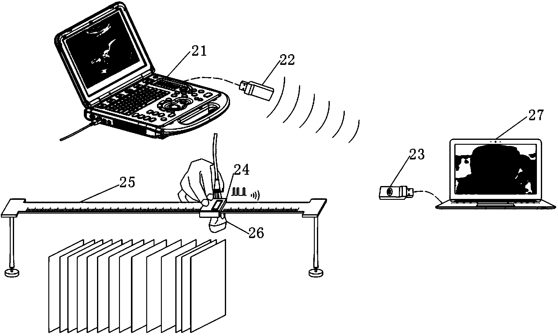
[0049] Present embodiment except following feature other structures are with embodiment 1: image 3 A schematic structural diagram of the system of this embodiment is given. The device described in this embodiment specifically includes: a server end, a client end 27 , a medical ultrasonic diagnostic instrument 21 , a bracket, a linear slide rail 25 , a slider 24 and an ultrasonic probe 26 .
[0050] Different from Embodiment 1, the second wireless transmission module in this embodiment only includes a USB wireless network card 22, and the third transmission module is a Bluetooth adapter 23, and the Bluetooth adapter 23 and the first wireless transmission module are connected through a wireless network for transmission For positioning information, the USB wireless network card 22 and the Bluetooth adapter 23 are connected through a wireless network for transmitting the intercepted two-dimensional ultrasound images.
[0051] The steps of the wireless three-dimensional ultrasonic...
Embodiment 3
[0058] Such as Figure 4 As shown, the device described in this embodiment specifically includes: a server end, a client end 34 , a medical ultrasonic diagnostic instrument 31 , and an ultrasonic probe 33 . The server end is provided with a first wireless transmission module 32, which is used to intercept several consecutive frames of two-dimensional ultrasound images displayed by the medical ultrasonic diagnostic instrument 31, and sends the images to the client terminal 34 through the first wireless transmission module 32; the client terminal 34 is provided with The second wireless transmission module is used for receiving images. After receiving the image, the client performs image processing and three-dimensional reconstruction. This embodiment adopts the free-arm scanning mode, which does not require brackets, linear slide rails and positioning devices.
[0059] The steps of the wireless three-dimensional ultrasonic imaging method in this embodiment are as follows:
[...
PUM
 Abstract
Description
Claims
Application Information
 - R&D
- Intellectual Property
- Life Sciences
- Materials
- Tech Scout
- Unparalleled Data Quality
- Higher Quality Content
- 60% Fewer Hallucinations
Browse by: Latest US Patents, China's latest patents, Technical Efficacy Thesaurus, Application Domain, Technology Topic, Popular Technical Reports.
© 2025 PatSnap. All rights reserved.Legal|Privacy policy|Modern Slavery Act Transparency Statement|Sitemap|About US| Contact US: help@patsnap.com


