Method for 3D printing of head and brain models with multiple materials at low cost
A 3D printing, multi-material technology, applied in the field of brain medical model manufacturing, can solve the problems of complex operation, increase the cost of medical model production, high price, etc., and achieve the effect of reducing costs
- Summary
- Abstract
- Description
- Claims
- Application Information
AI Technical Summary
Problems solved by technology
Method used
Image
Examples
Embodiment 1
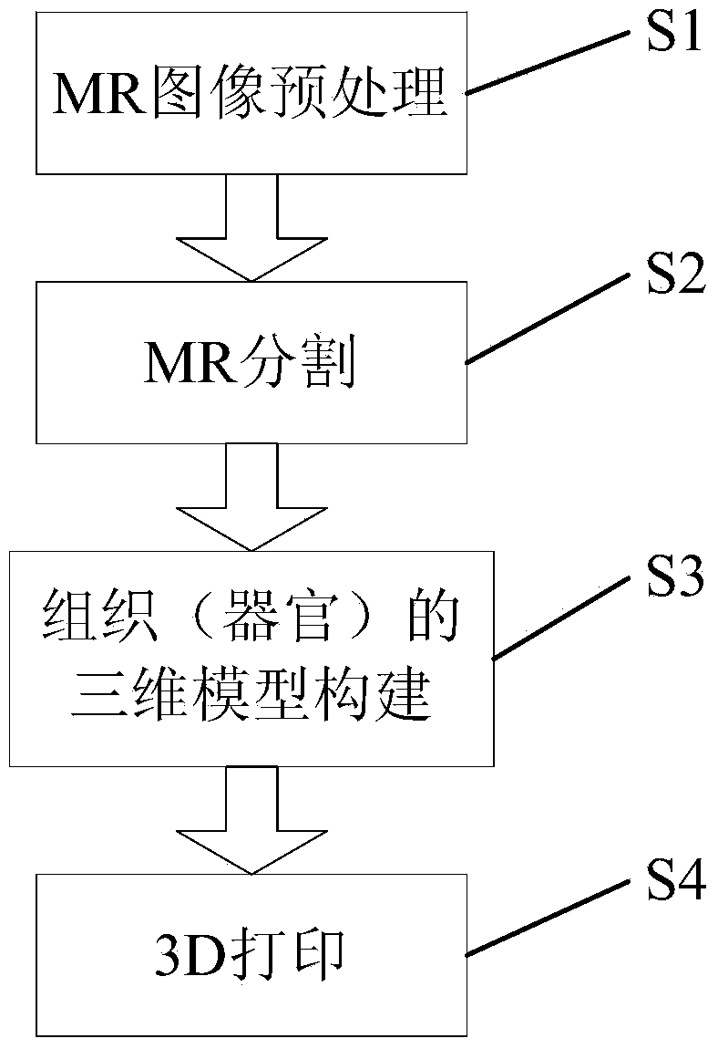
[0055] A method based on low-cost 3D printing of a multi-material head medical model, the specific implementation steps of the method are as follows.
[0056] MR image preprocessing:
[0057] Normalize the MR images by using reorientation and resampling algorithms to obtain images with a size of 256×256×256;
[0058] The non-uniform gray-scale correction algorithm is used to correct the gray-scale of the MR image.
[0059] Segmentation of MR images
[0060] The Marching Cubes algorithm is used to construct the initial three-dimensional surface of brain tissue for brain MR images, and the three-dimensional surface is smoothed by the spatial smoothing force to obtain evenly spaced vertices; then, the brain tissue and non-brain tissue are distinguished by the driving force of the gray scale of the image, and then , the vertices are driven by the brain probability map to move to the real brain tissue boundary, and finally the brain tissue without the skull and skin is obtained. ...
Embodiment 2
[0079] In this embodiment, the steps of MR image preprocessing, MR image segmentation and tissue (organ) three-dimensional model establishment are the same as in Embodiment 1. The difference from Embodiment 1 lies in the 3D printing process. The specific steps are as follows :
[0080] For brain tissue, set the order of printing 90 brain regions one by one in the order of brain regions;
[0081] Slice the 3D mesh model, set the slice thickness, and convert the 3D model into layered cross-sectional data.
[0082] Choose calcium phosphate as the skull printing material, choose medical gelatin to print the soft tissues of the skin and brain, and color suitable colors for different soft tissues;
[0083] Set the layer thickness of 3D printing to 1mm;
[0084] After setting the printing speed, feeding speed and other parameters, the upper computer is connected to the 3D printer for printing: first, print the white matter of a certain brain area, and then print the gray matter of ...
Embodiment 3
[0087] In this embodiment, the steps of MR image preprocessing, MR image segmentation, and establishment of three-dimensional models of tissues (organs) are the same as those in this embodiment. The difference from Embodiments 1 and 2 lies in the 3D printing process. The specific steps are as follows:
[0088] Slice the 3D surface model, set the slice thickness, and convert the 3D model into layered cross-sectional data.
[0089] Choose calcium phosphate as the skull printing material, choose medical gelatin to print the soft tissues of the skin and brain, and color suitable colors for different soft tissues;
[0090] Set the layer thickness of 3D printing to 1mm;
[0091] After setting the printing speed, feeding speed and other parameters, the upper computer is connected to the 3D printer for printing: first, print the white matter as a whole; then, set the filling rate and supports of the gray matter; then, print the outer surface of the gray matter, and the printing thick...
PUM
 Abstract
Description
Claims
Application Information
 - R&D
- Intellectual Property
- Life Sciences
- Materials
- Tech Scout
- Unparalleled Data Quality
- Higher Quality Content
- 60% Fewer Hallucinations
Browse by: Latest US Patents, China's latest patents, Technical Efficacy Thesaurus, Application Domain, Technology Topic, Popular Technical Reports.
© 2025 PatSnap. All rights reserved.Legal|Privacy policy|Modern Slavery Act Transparency Statement|Sitemap|About US| Contact US: help@patsnap.com


