Three-dimensional foramen intervertebral lens

A three-dimensional technology of intervertebral foramenoscopy, applied in the field of intervertebral foramenoscopy, can solve the problems of poor precision imaging of intelligent robots, inability of surgeons to be complicated and delicate, and affect the effect of surgery, so as to restore visual advantages and overcome the steep learning curve , The effect of easy operation

Inactive Publication Date: 2014-09-03
THE THIRD AFFILIATED HOSPITAL OF SUN YAT SEN UNIV
View PDF6 Cites 5 Cited by
  • Summary
  • Abstract
  • Description
  • Claims
  • Application Information

AI Technical Summary

Problems solved by technology

[0003] However, at present, the difficulty faced by the wider promotion of transforaminal endoscopic surgery is mainly that the learning curve for mastering this technique is very steep, requiring sufficient patience and experience, especially for beginners, which often affects the surgical effect; in addition, intelligence Robotic technology has been applied

Method used


Image

  • Three-dimensional foramen intervertebral lens
  • Three-dimensional foramen intervertebral lens

Examples


Embodiment Construction

[0016] The present invention will be further elaborated below in conjunction with accompanying drawing, wherein, the direction of the present invention is with figure 1 as standard.

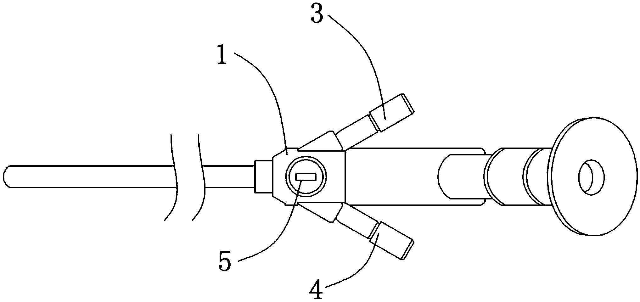
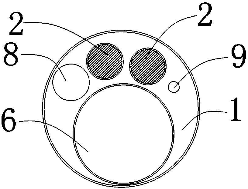
[0017] like figure 1 As shown, the three-dimensional intervertebral foramen mirror of the present invention includes a mirror tube body 1, a camera assembly and an imaging channel 2, a water inlet pipe 3, a water outlet pipe 4, and a lighting tube 5, wherein the camera assembly and the imaging channel 2 are fixedly installed on the mirror tube body 1, use the camera assembly and imaging channel 2 to obtain real-time stereoscopic image signals corresponding to the insertion end (left end) of the mirror tube body 1 in the intervertebral disc, and transmit the acquired stereoscopic image signals to an external imaging device connected to it for further processing. Three-dimensional imaging and display; the water inlet pipe 3 and the water outlet pipe 4 are respectively fixedly installed on the side...


PUM


Abstract

The invention discloses a three-dimensional foramen intervertebral lens which is simple in structure. A surgical operation position can be accurately located. An operator can carry out learning and operation conveniently. The three-dimensional foramen intervertebral lens comprises a lens pipe body, a camera shooting component, imaging channels, a water inlet pipe, a water outlet pipe and a lighting pipe. An operation channel, the imaging channels, a water inlet channel and a water outlet channel which are independent are arranged on the lens pipe body, and left end openings of the channels are formed in the left end of the lens pipe body. The middle portions of the imaging channels are communicated with the lighting pipe. Another opening of the water inlet channel is communicated with a water inlet pipe. Another opening of the water outlet channel is communicated with the water outlet pipe. The water inlet pipe, the water outlet pipe and the lighting pipe are fixedly connected with the side wall of the lens pipe body. The camera shooting component and the imaging channels further comprise two three-dimensional endoscopes and two optical fibers. The two three-dimensional endoscopes are fixedly arranged at the left end openings of the two imaging channels respectively. The two optical fibers are arranged in the two imaging channels respectively. One end of each optical fiber is connected with a corresponding three-dimensional endoscope. The other ends of the optical fibers extend out of right side openings of the imaging channels.

Description

technical field [0001] The invention relates to an intervertebral foramina mirror used in spinal surgery, in particular to a three-dimensional intervertebral foramina mirror with simple structure, which can precisely locate the operation position and facilitate the learning and operation of the operator. Background technique [0002] The current development direction of spinal surgery technology is minimally invasive and intelligent. On the premise of achieving the same or better surgical effect than traditional open surgery, surgical trauma should be reduced as much as possible. The application of transforaminal endoscopic surgery in the treatment of lumbar spine diseases has been widely recognized in spinal surgery. Current research shows that lumbar diseases that required traditional classic discectomy in the past are suitable for transforaminal endoscopic surgery. Both methods have achieved good results. The effect of the two operations is comparable, and the recurrence ...

Claims


Application Information

Patent Timeline
IPC IPC(8): A61B1/313A61B1/05A61B1/07A61B1/012
Inventor 戎利民舒涛董健文刘斌
Owner THE THIRD AFFILIATED HOSPITAL OF SUN YAT SEN UNIV
Why Eureka
Try Eureka