Electrocardio ultrasonic signal fusion computed tomography imaging system and method

A tomography, ultrasonic signal technology, applied in diagnosis, echo tomography, diagnostic recording/measurement, etc., can solve problems such as lack of electro-mechanical signals

Inactive Publication Date: 2014-10-01
镇郁琼 +1
View PDF7 Cites 15 Cited by
  • Summary
  • Abstract
  • Description
  • Claims
  • Application Information

AI Technical Summary

Problems solved by technology

Although one-dimensional electrocardiogram has been used in the field of clinical diagnosis in two-dimensional plane and three-dimensional ultrasound, it is limited to simple ECG monitoring and synchronous detection of one-dimensional electrocardiogram in the process of ultrasound examination, and does not have synchronization in two-dimensional and three-dimensiona...

Method used


Image

  • Electrocardio ultrasonic signal fusion computed tomography imaging system and method
  • Electrocardio ultrasonic signal fusion computed tomography imaging system and method
  • Electrocardio ultrasonic signal fusion computed tomography imaging system and method

Examples


Embodiment Construction

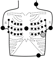
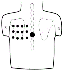
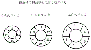
[0052] Explanation of terms: Cardiac anatomical tomography: that is, different anatomical layers from the apex to the base (such as Figure 4A , 4B The ECG signals and ultrasound signals of the anatomical parts of the left ventricle in 16 segments are acquired synchronously according to the anatomical structure).

[0053] The ECG signal scanning of different anatomical sections of the heart refers to the cardiac electrical activity of the two-dimensional cross-section of the heart, such as cardiac transmural potential, electrocardiogram, and cardiac activation maps such as depolarization and repolarization patterns. Its anatomical section positioning adopts the currently internationally accepted American Heart Association's cardiac anatomical section, which is a general section of international cardiac medical imaging technology, including ultrasound technology. This invention introduces this concept into ECG technology for the first time.

[0054] The present invention will...


PUM


Abstract

The invention relates to a medical examination and diagnosis method and instrument, in particular to a multi-dimensional electrocardio ultrasonic signal fusion computed tomography imaging system and method based on anatomical orientation. A multi-dimensional ultrasonic cardiac structure and a perfusion image technology are further integrated on the basis of a multi-dimensional electrocardiosignal imaging system and method, and a novel non-invasive platform is provided for recording and evaluating the electro-mechanical signal characteristic and myocardial perfusion characteristic of heart. The method is a unique electrocardio-mechanical function and perfusion synchronized computed tomography imaging method based on anatomical orientation, and the technology is a non-invasive imaging technology for detecting transmural cardiac electrical activity information and mechanical signals. By adopting the medical examination and diagnosis method and instrument, the diagnosis of myocardial diseases such as cardiac hypertrophy, cardiomyopathy, myocardial infarction and myocarditis, the determination of the origin of arrhythmia, and the like can be enhanced, and a brand-new diagnosis and guide treatment tool is provided for rapid real-time diagnosis of heart diseases.

Description

technical field [0001] The invention relates to a medical examination and diagnosis method and an instrument, in particular to a multi-dimensional electrocardiographic and ultrasonic signal fusion imaging system and method based on anatomical positioning. Background technique [0002] Cardiac electrical excitation and the resulting mechanical activity are the two most basic physiological features of the heart. The former reflects the self-discipline, excitability and electrical conductivity of the cardiac excitatory tissue, while the latter reflects the rhythmic contraction and relaxation of myocardial tissue to maintain the body's blood circulation. The electrical excitation and mechanical activity of the heart are closely related, which is called Electrical-Mechanical (E-M) Coupling in physiology, and this coupling is a necessary condition for ensuring the normal function of the heart. Many heart diseases can cause cardiac electrical activity and mechanical dysfunction. D...

Claims


Application Information

Patent Timeline
IPC IPC(8): A61B5/0402A61B8/13
Inventor 雷鸣朱天刚郝国梁
Owner 镇郁琼
Why Eureka
Try Eureka