
A method and device for image enhancement of electronic endoscope

An electronic endoscope and image enhancement technology, applied in image enhancement, image data processing, graphic image conversion and other directions, can solve the problems of blurred moving target imaging, unclear whole image, uneven image brightness, etc. Radiation interference, reduced lamp life, image enhancement effects

Active Publication Date: 2017-10-20
BEIJING HUACO HEALTHCARE TECH CO LTD
View PDF5 Cites 0 Cited by
  • Summary
  • Abstract
  • Description
  • Claims
  • Application Information

AI Technical Summary

Problems solved by technology

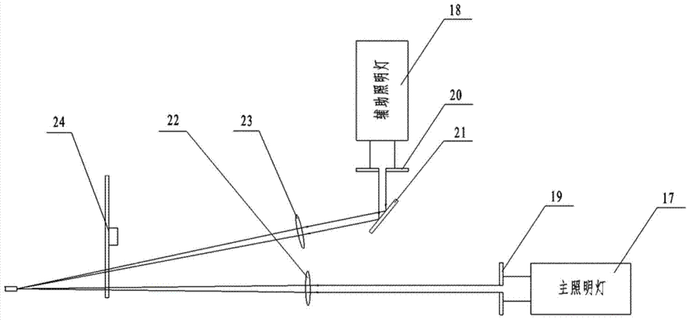
[0011] The purpose of the present invention is to solve the problem of uneven brightness of images collected by medical electronic endoscopes, to overcome the problem of partial unclearness of the entire image caused by the limited dynamic range of existing imaging devices, and to provide a method for improving the dynamic range of electronic endoscope images and changing the light source. The method of lighting brightness output, this method changes the light source output lighting brightness when two adjacent frames of images are imaged by the camera, and then synthesizes the two frames of images to achieve endoscopic high dynamic range image enhancement and overcome the moving target imaging caused by increasing the exposure time Vague

Method used


Image

  • A method and device for image enhancement of electronic endoscope
  • A method and device for image enhancement of electronic endoscope
  • A method and device for image enhancement of electronic endoscope

Examples


Embodiment Construction

[0021] Hereinafter, embodiments of the present invention will be described in detail with reference to the drawings.

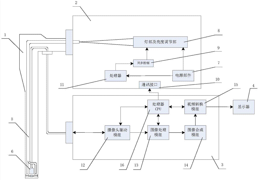
[0022] figure 1 It is a schematic composition diagram of an electronic endoscope system for improving the imaging dynamic range of an embodiment.

[0023] exist figure 1 Among them, the endoscope system includes: an endoscope mirror body 1, which has a light guide cable member 5 that guides the output light of the light source to the front end of the endoscope for illumination, and a micro camera 6 that photographs the inner cavity of the organ. The light source device 2 provides illumination light for the electronic endoscope system, and has a power supply part 7, which provides power for each part of the light source; the lamp part and the brightness adjustment part 8 provide illumination light with controllable brightness change to the light guide part 5 of the endoscope; The synchronous control part 9 is used to control the brightness adjustment of the l...


PUM


Abstract

The invention provides a method and device for image enhancement of an electronic endoscope. The method changes the output illumination brightness of the light source when two adjacent frames of the camera are imaged, and then synthesizes the two frames of images to realize the high dynamic range image of the endoscope. Enhanced to overcome the blurring of moving target imaging caused by increasing exposure time. According to the method of the present invention, there is provided an electronic endoscope device which realizes an improvement in the dynamic range of an image.

Description

technical field [0001] The invention relates to the technical field of medical electronic endoscope imaging, in particular to an electronic endoscope image enhancement method and device, and an electronic endoscope using the image enhancement device. Background technique [0002] In the medical field, an electronic endoscope system is used to check the internal lesions of organs. The composition of the endoscope system includes a light source device that emits illuminating light, a front end equipped with a camera unit, a flexible insertion mirror body with a light guide cable, and a camera unit. An image processing device for driving control and processing and enhancing the captured image, and a display for displaying the processed image. [0003] During the imaging process of the endoscope, due to the influence of factors such as the relative movement of the camera device and the inspected organ, electronic noise, interference, etc., the obtained endoscopic image will inev...

Claims


Application Information

Patent Timeline
Patent Type & Authority Patents(China)
IPC IPC(8): G06T5/50G06T3/40A61B1/00
Inventor 周智峰段海峰杨云生邹慧玲
Owner BEIJING HUACO HEALTHCARE TECH CO LTD
Learn More