
Medical imaging system having a magnetic resonance imaging unit and a positron emission tomography (PET) unit

A medical imaging system and magnetic resonance imaging technology, which is applied in the fields of magnetic resonance measurement, medical science, and magnetic variable measurement, can solve the problems of inability to collect data in parallel, deteriorate the quality of magnetic resonance image data, and increase the complexity of the inspection process. Achieve the effect of saving space and compact arrangement

Inactive Publication Date: 2014-11-12
SIEMENS AG
View PDF7 Cites 4 Cited by
  • Summary
  • Abstract
  • Description
  • Claims
  • Application Information

AI Technical Summary

Problems solved by technology

In addition, the use of the local radio-frequency antenna unit increases the complexity of the examination procedure of the hybrid imaging examination to be carried out including PET examination and magnetic resonance examination, because the local radio-frequency antenna unit also has to be positioned on the patient before the imaging examination.
[0004] In order to eliminate this problem, it is possible to dispense with the use of local high-frequency antenna elements during the hybrid imaging examination, however, this significantly deteriorates the quality of the magnetic resonance image data
In addition, the examination duration is increased accordingly, since the data cannot be acquired in parallel

Method used


Image

  • Medical imaging system having a magnetic resonance imaging unit and a positron emission tomography (PET) unit
  • Medical imaging system having a magnetic resonance imaging unit and a positron emission tomography (PET) unit
  • Medical imaging system having a magnetic resonance imaging unit and a positron emission tomography (PET) unit

Examples


Embodiment Construction

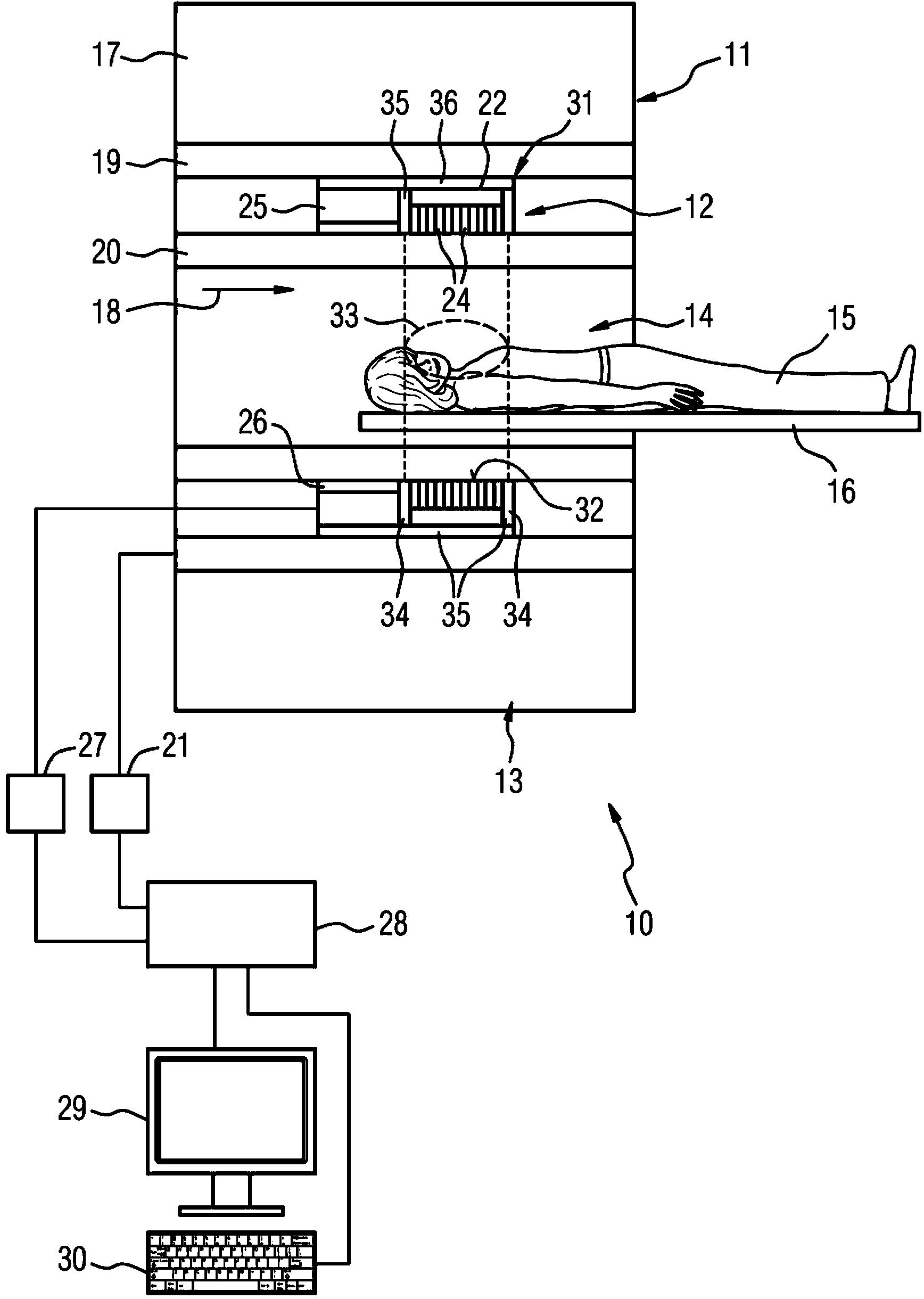
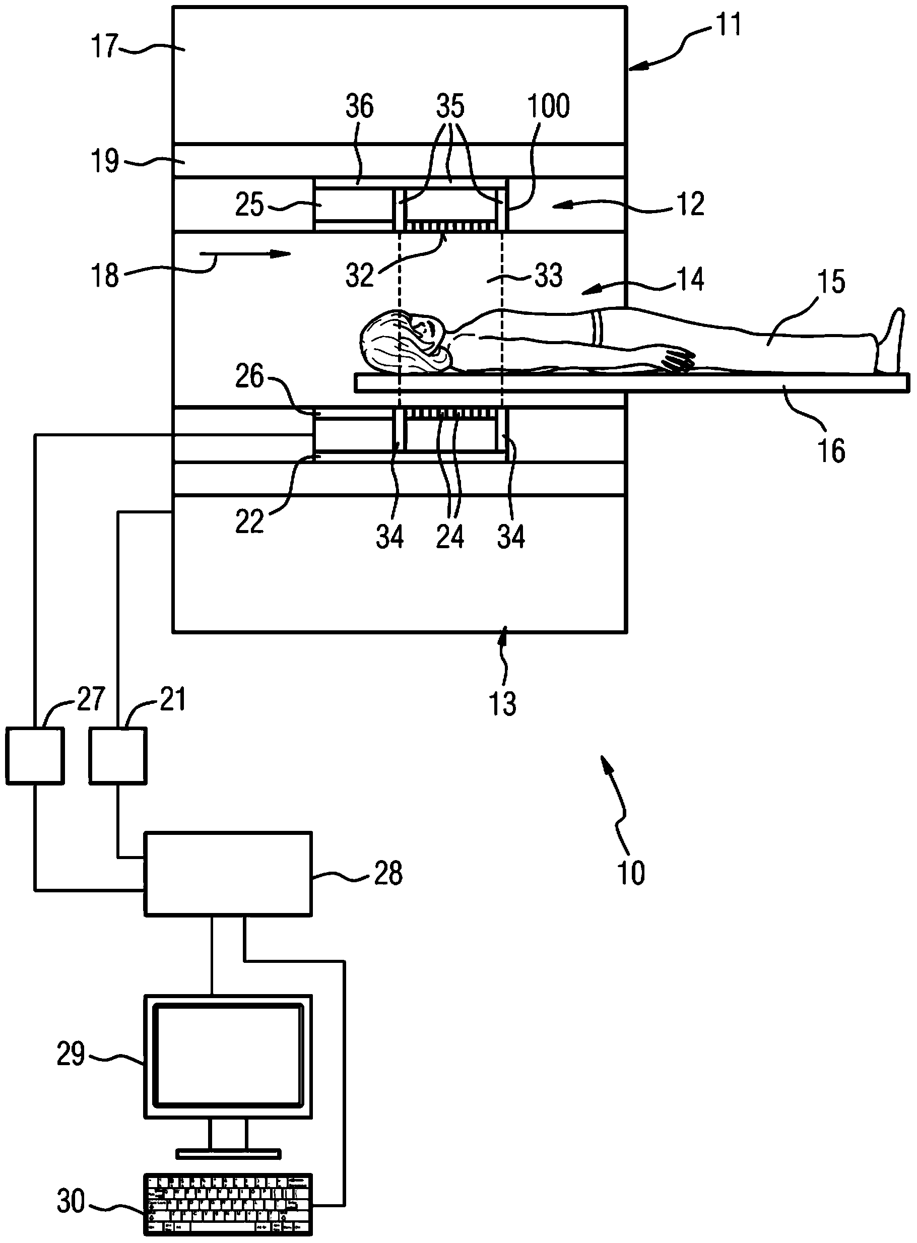
[0024] exist figure 1 A medical imaging system 10 is shown in FIG. The medical imaging system 10 is constituted by a combined imaging system including a magnetic resonance imaging unit 11 and a positron emission tomography unit 12 (PET unit 12 ).

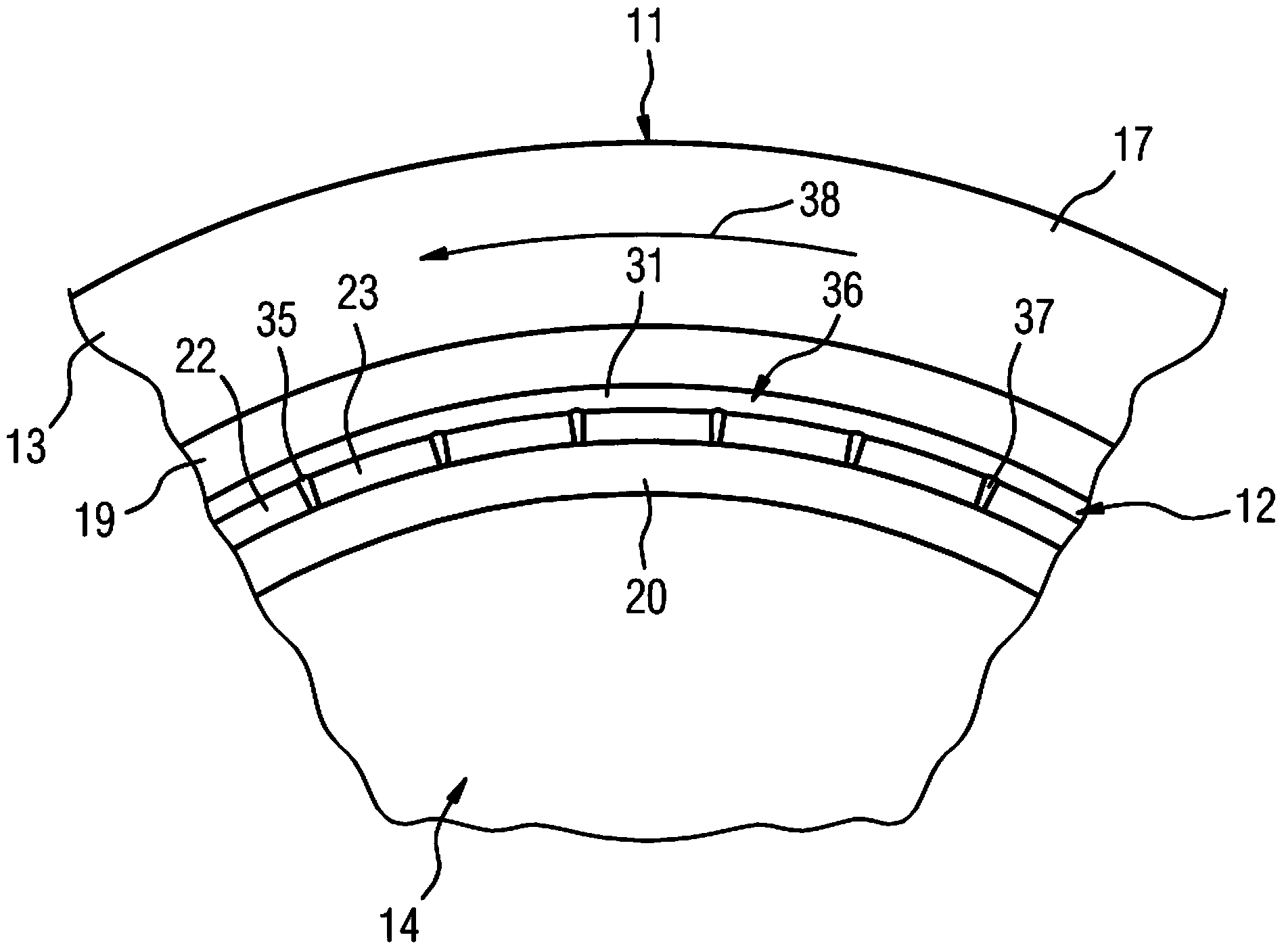
[0025]The magnetic resonance imaging unit 11 comprises a magnet unit 13 and a patient receiving space 14 surrounded by the magnet unit 13 for receiving a patient 15 , wherein the patient receiving space 14 is surrounded in the circumferential direction 38 by a magnet unit (not shown in detail) surrounded by the magnet unit 13 . The housing partition unit of the resonance imaging unit 11 surrounds it cylindrically. The patient 15 can be moved into the patient receiving space 14 by means of the patient support device 16 of the magnetic resonance imaging unit 11 . For this purpose, the patient support device 16 is arranged displaceably within the patient receiving space 14 .

[0026] The magnet unit 13 includes a main magnet 17 whic...


PUM


Abstract

A medical imaging system includes a magnetic resonance imaging unit including a magnet unit and a patient receiving area surrounded by the magnet unit, wherein the magnet unit includes a main magnet, a gradient coil unit and a radio-frequency antenna unit. In at least one embodiment, the medical imaging system further includes a positron emission tomography unit including at least two positron emission tomography detector modules. The at least two positron emission tomography detector modules each include a detector surface. Further, the radio-frequency antenna unit is disposed outside an area disposed between an examination area and the detector surfaces.

Description

technical field [0001] The invention relates to a medical imaging system having a magnetic resonance imaging unit and a positron emission tomography (PET) unit comprising a magnet unit and a patient accommodation space surrounded by the magnet unit, wherein the magnet unit comprises a main magnet, a gradient coil unit and A radio-frequency antenna unit, wherein the positron emission tomography unit has at least two positron emission tomography detector modules. Background technique [0002] In medical imaging systems comprising a magnetic resonance imaging unit and a positron emission tomography unit (PET unit), magnetic resonance image data are often acquired by means of a local high-frequency antenna unit. The partial radio-frequency antenna unit is preferably arranged around a partial region of the patient to be examined, for example a partial head antenna unit surrounds the patient's head and / or a partial body antenna unit surrounds the patient's torso region, etc. Duri...

Claims


Application Information

Patent Timeline
IPC IPC(8): A61B5/055A61B6/03
CPCG01R33/481G01R33/34
Inventor M.芬彻尔B.海斯曼K.加特克R.雷德贝克S.施米特
Owner SIEMENS AG
Learn More