Method for extracting spinal marrow in CT (Computed Tomography) image

A CT image and extraction method technology, applied in the field of image processing, can solve complex and computationally intensive problems

Active Publication Date: 2015-02-11
SHANGHAI UNITED IMAGING HEALTHCARE
View PDF3 Cites 15 Cited by
  • Summary
  • Abstract
  • Description
  • Claims
  • Application Information

AI Technical Summary

Problems solved by technology

However, this method is too complex ...

Method used


Image

  • Method for extracting spinal marrow in CT (Computed Tomography) image
  • Method for extracting spinal marrow in CT (Computed Tomography) image
  • Method for extracting spinal marrow in CT (Computed Tomography) image

Examples


Embodiment Construction

[0054] In the following description, numerous specific details are set forth in order to provide a thorough understanding of the present invention. However, the present invention can be implemented in many other ways different from those described here, and those skilled in the art can make similar extensions without violating the connotation of the present invention, so the present invention is not limited by the specific implementations disclosed below.

[0055] Secondly, the present invention is described in detail by means of schematic diagrams. When describing the embodiments of the present invention in detail, for convenience of explanation, the schematic diagrams are only examples, which should not limit the protection scope of the present invention.

[0056] A method for extracting the spinal cord in a CT image of the present invention will be described in detail below with reference to the drawings and embodiments. The method for extracting the spinal cord in the CT i...


PUM


Abstract

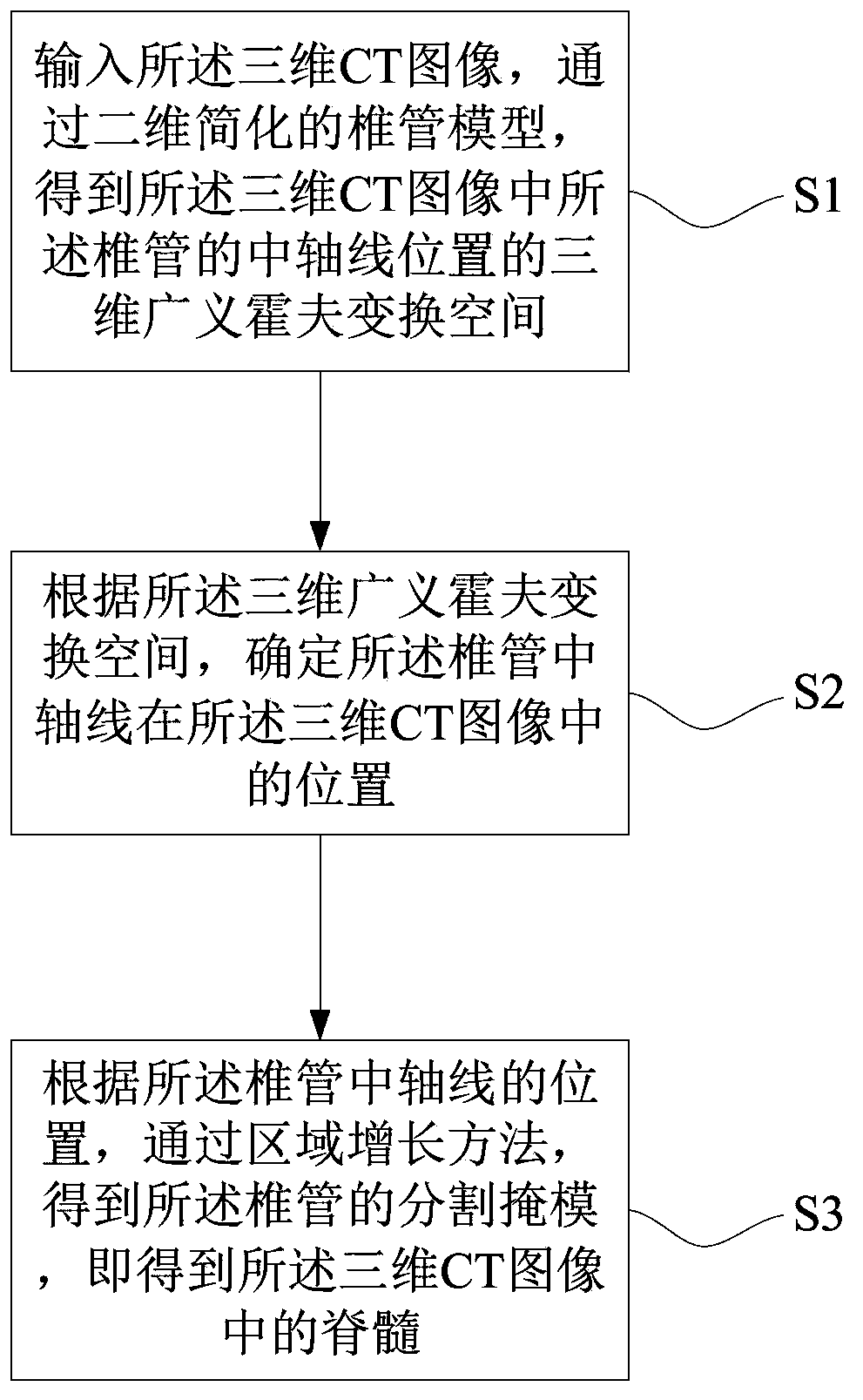
The invention provides a method for extracting spinal marrow in a CT (Computed Tomography) image. The method comprises the following steps: (1) inputting the three-dimensional CT image and obtaining a three-dimensional GHT (Generalized Hough Transform) space of the position of a central axis of a spinal canal in the three-dimensional CT image by a two-dimensional simplified spinal canal model; (2) according to the three-dimensional GHT space, determining the position of the central axis of the spinal canal in the three-dimensional CT image; (3) according to the position of the central axis of the spinal canal, obtaining a dividing mask of the spinal canal, i.e. obtaining the spinal marrow in the three-dimensional CT image, by a region growing method. According to the technical scheme, not only can the spinal marrow in the CT image be automatically and rapidly extracted, but also the method has higher stability and flexibility.

Description

technical field [0001] The invention relates to the field of image processing, in particular to a method for extracting spinal cord in CT images. Background technique [0002] In the preparation stage of radiotherapy, the doctor calculates the radiation route and dose used in the radiotherapy process through the patient's CT image, and outlines (segments) some important organs in the CT image at the same time, so that it can avoid the radiation radiation during radiotherapy. irradiated, or reduced radiation doses received. The spinal cord is a core component of the nervous system and an important area that must be protected during radiation therapy. When planning radiation therapy for cancers of the head (such as brain, neck), chest (such as breast, lung, esophagus), and abdomen (such as liver, spleen, kidney, pancreas), doctors need to protect the spinal cord. Segmented from pre-acquired CT images. [0003] At present, the automatic segmentation of spinal canal in 3D CT ...

Claims


Application Information

Patent Timeline
IPC IPC(8): G06T7/00G06K9/46A61B6/03
CPCA61B6/03A61B6/5229G06T7/12G06T2207/30012
Inventor 刘靖李强
Owner SHANGHAI UNITED IMAGING HEALTHCARE
Why Eureka
Try Eureka