Three-dimensional ultrasonic imaging method and system

A technology of three-dimensional ultrasound and imaging methods, which is applied in ultrasound/acoustic/infrasonic diagnosis, image enhancement, image analysis, etc., and can solve problems such as lack of understanding of three-dimensional space

Active Publication Date: 2015-03-18
SHENZHEN MINDRAY BIO MEDICAL ELECTRONICS CO LTD +1
View PDF4 Cites 32 Cited by
  • Summary
  • Abstract
  • Description
  • Claims
  • Application Information

AI Technical Summary

Problems solved by technology

However, doctors need to have a very deep understanding of three-dimensional space in order to be able to adjust the median sagittal pl...

Method used


Image

  • Three-dimensional ultrasonic imaging method and system
  • Three-dimensional ultrasonic imaging method and system
  • Three-dimensional ultrasonic imaging method and system

Examples


Embodiment Construction

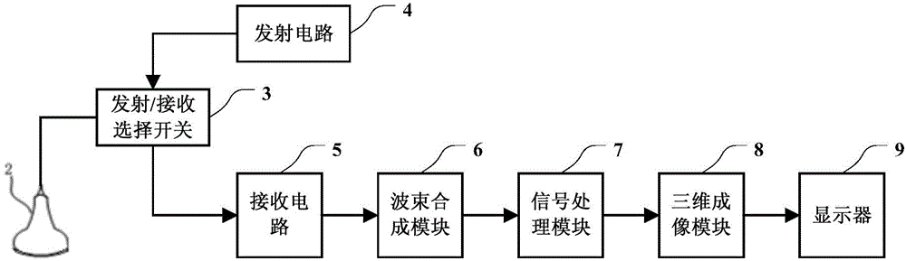
[0038] Such as figure 1 Shown is the structural block diagram of the three-dimensional ultrasound imaging system. The three-dimensional ultrasonic imaging system includes a probe 2 , a transmit / receive selection switch 3 , a transmit circuit 4 , a receive circuit 5 , a beam forming module 6 , a signal processing module 7 , a three-dimensional imaging module 8 , and a display 9 . Transmitting circuit 4 sends a group of delayed focused pulses to probe 2, and probe 2 transmits ultrasonic waves to the body tissue under test (not shown in the figure), and receives the tissue-bearing tissue reflected from the body tissue under test after a certain delay. The ultrasonic echo of the information is converted back into an electrical signal. The receiving circuit 5 receives these electrical signals, and sends these ultrasonic echo signals to the beamforming module 6 . The ultrasonic echo signal completes focus delay, weighting and channel summation in the beamforming module 6 , and the...


PUM


Abstract

A three-dimensional ultrasonic imaging method and a three-dimensional ultrasonic imaging system. The method comprises: transmitting an ultrasonic wave to a fetal head; receiving an ultrasonic echo, obtaining an ultrasonic echo signal, and obtaining the three-dimensional volume data of the fetal head according to the ultrasonic echo signal; according to the characteristics of a median sagittal section of the fetal head, detecting the median sagittal section in the three-dimensional volume data; and displaying the median sagittal section. The ultrasonic imaging method and the ultrasonic imaging system can obtain the three-dimensional volume data of the fetal head by ultrasonically scanning the fetus and can also automatically detect and display the median sagittal section of the fetal head, thus solving the problem that it is difficult for a doctor to precisely and manually position the median sagittal section.

Description

technical field [0001] The invention relates to the technical field of medical ultrasound imaging, in particular to a three-dimensional ultrasound imaging method and system. Background technique [0002] Ultrasound instruments are generally used by doctors to observe the internal tissue structure of the human body. The doctor places the ultrasonic probe on the skin surface corresponding to the part of the human body to obtain an ultrasonic image of the part. Ultrasound has become one of the main auxiliary methods for doctors' diagnosis due to its safety, convenience, non-destructive, and low-cost characteristics. [0003] Obstetrics is one of the most widely used fields of ultrasound diagnosis. In this field, ultrasound avoids the influence of X-rays and other maternal and fetal effects, and its application value is obviously superior to other imaging examination equipment. Ultrasound can not only observe and measure fetal morphology, but also obtain various physiological a...

Claims


Application Information

Patent Timeline
IPC IPC(8): A61B8/00
CPCA61B8/00A61B8/14A61B8/0808A61B8/0866A61B8/4444A61B8/466A61B8/483A61B8/523A61B8/469A61B8/5223G06T2207/30044G06T2207/10136G06T7/68A61B8/461A61B8/5207
Inventor 邹耀贤林穆清陈志杰熊奕姚斌
Owner SHENZHEN MINDRAY BIO MEDICAL ELECTRONICS CO LTD
Why Eureka
Try Eureka