Anterior cervical and craniocervical fixing device

A fixation device, anterior approach technology, applied in the direction of fixator, internal fixator, internal bone synthesis, etc., to achieve the effect of reducing operation time, reducing operation risk, and improving stability

Inactive Publication Date: 2015-03-25
NANFANG HOSPITAL OF SOUTHERN MEDICAL UNIV
View PDF9 Cites 26 Cited by
  • Summary
  • Abstract
  • Description
  • Claims
  • Application Information

AI Technical Summary

Problems solved by technology

However, there is no specific fixation device fo

Method used


Image

  • Anterior cervical and craniocervical fixing device
  • Anterior cervical and craniocervical fixing device
  • Anterior cervical and craniocervical fixing device

Examples


Embodiment 1

[0023] Embodiment 1: Anterior cervical craniocervical fixation device according to the present invention

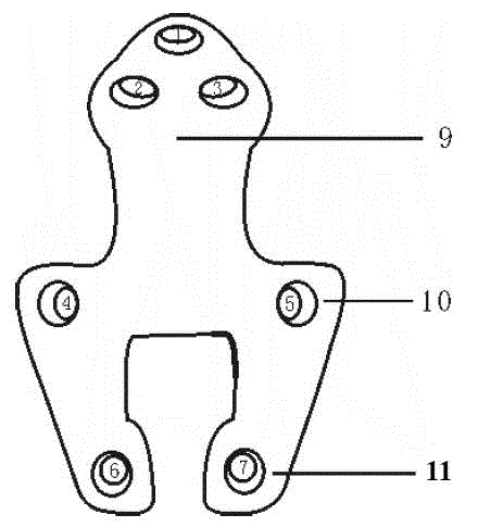
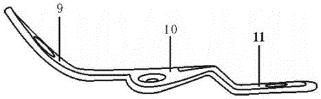
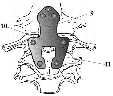
[0024] Anterior craniocervical fixation device of the present invention, such as figure 1 and figure 2 As shown, it includes: a section of inclined slope fixing section 9 and a section of horizontal atlas fixing section 10; and an axis fixing section 11 extending downward from both sides of the atlas fixing section. The slope fixing part 9 and the atlas fixing part 10 form a certain angle so that the slope fixing part is suitable to be close to the anterior slope of the human neck and the axis vertebrae fixing part is suitable to be close to the human cervical 2 vertebral body. Preferably, the angle between the slope fixing part 9 and the atlas fixing part 10 is 114° to 148°.

[0025] Such as figure 1 As shown, three screw holes 1, 2 and 3 are arranged on the slope fixing part 9, and these screw holes are arranged in a triangle, so that the screws can pass through the...

Embodiment 2

[0035] Embodiment 2: The improved anterior cervical craniocervical fixation device according to the present invention

[0036] The improved anterior cervical craniocervical fixation device of the present invention, such as Figure 5 and Figure 6 As shown, it includes: a section of inclined slope fixation part 9 and a section of horizontal atlas fixation part 10; and an axis fixation part 11 extending downward from the atlas fixation part. The slope fixing part 9 and the atlas fixing part 10 form a certain angle so that the slope fixing part is suitable for being close to the anterior slope of the human neck, and the axis vertebra fixing part is suitable for being close to the human cervical 2 vertebral body. When the atlas fixation part 10 is extended to form the axis fixation part 11, an open groove 12 is opened in the center, so that after being fixed by the improved anterior cervical craniocervical fixation device, the rear joint space can be passed through the groove. P...

Embodiment 3

[0048] Example 3: Biomechanical Evaluation of the Stability of the Anterior Cervical Craniocervical Fixation Device

[0049] 1. Experimental purpose

[0050] From the perspective of biomechanics, the stability of the anterior craniocervical fixation device of the present invention for upper cervical spine fixation is discussed.

[0051] 2. Experimental materials

[0052] Anterior craniocervical fixation device: it is made of a steel plate, and its structure is referred to in Example 1, hereinafter referred to as "anterior craniocervical fixation plate".

[0053] The occipital-cervical fixation device, hereinafter referred to as "posterior occipital-cervical fixation device", is different from "anterior craniocervical fixation plate".

[0054] Kirschner wire.

[0055] Screws (3.5mm diameter).

[0056] Six cadaveric head and neck specimens (from occipital condyle C0 to C3 vertebrae, 4 males, 2 females; mean age, 46 years; range 35–62 years).

[0057] 3. Experimental method ...


PUM

PropertyMeasurementUnit
Diameteraaaaaaaaaa
Diameteraaaaaaaaaa

Abstract

The invention relates to an anterior cervical and craniocervical fixing device which is of an integrally-formed three-section structure. The anterior cervical and craniocervical fixing device comprises a front section of a clivus fixing part, a middle section of an atlas fixing part and a rear section of a dentate fixing part. The front part of the clivus fixing part is oblique, a certain angle is formed between the clivus fixing part and the atlas fixing part, and thus the clivus fixing part is suitable for being tightly attached to anterior cervical clivus of a human body. The atlas fixing part is horizontal, extends to the two sides and then extends backwards to form the dentate fixing part. The axis fixing part extends on the rear part of the atlas fixing part, and thus the dentate fixing part is suitable for being tightly attached to the cervical 2 vertebral body of the human body. The fixing device special for fixing anterior cervical and craniocervical is proposed the first time, better stability is achieved, the anterior cervical and the craniocervical can be fixed together, body turnover for fixing posterior occipitocervical is not needed after anterior release and pressure reduction are conducted in the operation process of a patient, the operation risk is greatly reduced, the operation time is shortened, and recovery of the patient is facilitated.

Description

technical field [0001] The invention relates to an orthopedic clinical instrument, in particular to an anterior cervical craniocervical fixation device. Background technique [0002] Clinically, various types of craniospinal junction disorders are common. Due to the complex anatomical structure and adjacent to important nerves and blood vessels, it is difficult to treat and is a difficult point in spinal surgery. For severe skull basilar depression with atlanto-occipital ossification, irreversible atlantoaxial dislocation, atlantoaxial bony fusion dislocation, upward movement of the odontoid process into the foramen magnum and other causes of spinal cord ventral compression, often need Release and reset through the anterior cervical approach or through the mouth, or directly remove various direct compressive objects such as hyperplastic granulation tissue, scar and callus, and indirectly or directly relieve the compressive factors on the ventral side of the spinal cord, and...

Claims


Application Information

Patent Timeline
IPC IPC(8): A61B17/80A61B17/70
CPCA61B17/7059A61B17/8061
Inventor 朱青安季伟陈建庭孔刚刚吴秀华
Owner NANFANG HOSPITAL OF SOUTHERN MEDICAL UNIV
Why Eureka
Try Eureka