
Three-dimensional imaging flow cytometer device

A flow cytometer and three-dimensional imaging technology, applied in the field of optical instruments, can solve the problems of affecting life, affecting the efficiency of flow test, slow flow rate, etc., and achieving the effect of good authenticity

Inactive Publication Date: 2015-04-08
CHANGCHUN INST OF OPTICS FINE MECHANICS & PHYSICS CHINESE ACAD OF SCI
View PDF10 Cites 16 Cited by
  • Summary
  • Abstract
  • Description
  • Claims
  • Application Information

AI Technical Summary

Problems solved by technology

[0005] The present invention solves the problem that the existing imaging flow cytometer device scans the cells due to the slow liquid flow speed, which affects the efficiency of the flow test
In addition, the sample flow needs to be in direct contact with the microscope objective lens, and the sample flow can easily pollute the microscope objective lens and affect its life. A three-dimensional imaging flow cytometer is provided

Method used


Image

  • Three-dimensional imaging flow cytometer device
  • Three-dimensional imaging flow cytometer device

Examples


specific Embodiment approach 1

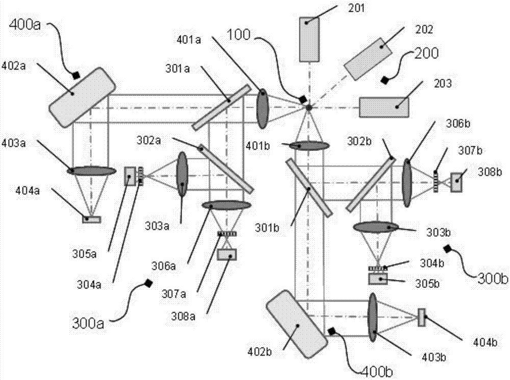
[0012] Specific implementation mode 1. Combination figure 1 with figure 2 In this embodiment, the three-dimensional imaging flow cytometer device uses two sets of imaging flow cytometry systems to simultaneously image the moving sample in two directions to obtain the final three-dimensional image; the device includes a sample injection unit 100, a light source 200. Two sets of speed measurement-focus units and two sets of imaging units; the structures and working principles of the two imaging flow systems are identical, and each imaging flow system includes a speed measurement-focus unit and an imaging unit;

[0013] The sample injection unit 100 ensures that samples such as viruses, cells, microspheres or small model organisms pass through the imaging detection area individually and side by side at high speed.

[0014] The light source 200 includes a first light source 201, a second light source 202 and a third light source 203. The first light source 202 emits side scatter...


PUM


Abstract

The invention relates to a three-dimensional imaging flow cytometer device, relating to the field of biological and medical optical apparatuses and aiming at solving the problems that the flow testing efficiency is influenced by low speed of a liquid flow when cells are scanned by an existing imaging flow cytometer device and a micro-objective is easily polluted by a sample flow and the service life of the micro-objective is influenced since the sample flow needs to directly contact the micro-objective, etc. A moving cell is synchronously imaged in two directions through two vertical imaging flow systems; the relative position distribution of the internal structure of the cell is extracted; and therefore, the three-dimensional structural image of the cell is obtained. A lateral scattering and fluorescence exciting laser light source illuminates at 45 DEG and is shared by a speed-measuring and focusing unit and an imaging unit. By means of three-dimensional cell imaging, the original position information of the internal structure of the cell is kept; the morphological description authenticity is better; and thus, the imaging flow cytometer is capable of obtaining more meaningful biomedical information.

Description

technical field [0001] The invention relates to the field of biological and medical optical instruments, in particular to a three-dimensional imaging flow cytometer device. Background technique [0002] Flow cytometry is a technique for rapid quantitative analysis and sorting of cells or other biological particles (such as microspheres, bacteria, small model organisms, etc.) arranged in a single row in a liquid flow. In the fields of biology and medicine, when a large number of cells need to be scanned, the flow cytometer completely sacrifices the spatial resolution to obtain a high detection speed, tens of thousands of cells per second. Imaging flow cytometry can not only obtain population analysis data of a large number of cells, but also can see cell images in real time, and the analysis results of each step can be confirmed by images. Compared with traditional flow cytometers, imaging flow cytometers have greater advantages when it is necessary to obtain cell morphology...

Claims


Application Information

Patent Timeline
IPC IPC(8): G01N15/14
Inventor 刘英李灿李淳王健郭帮辉张建忠孙强赵建
Owner CHANGCHUN INST OF OPTICS FINE MECHANICS & PHYSICS CHINESE ACAD OF SCI
Learn More