

Kit for detecting protein glycosylation subtype and detection method thereof
A detection kit and a technique for isolating isoforms are applied in the field of protein and post-translational modification detection to achieve the effect of improving detection efficiency and wide applicability
- Summary
- Abstract
- Description
- Claims
- Application Information
AI Technical Summary
Problems solved by technology
Method used
Image

Examples
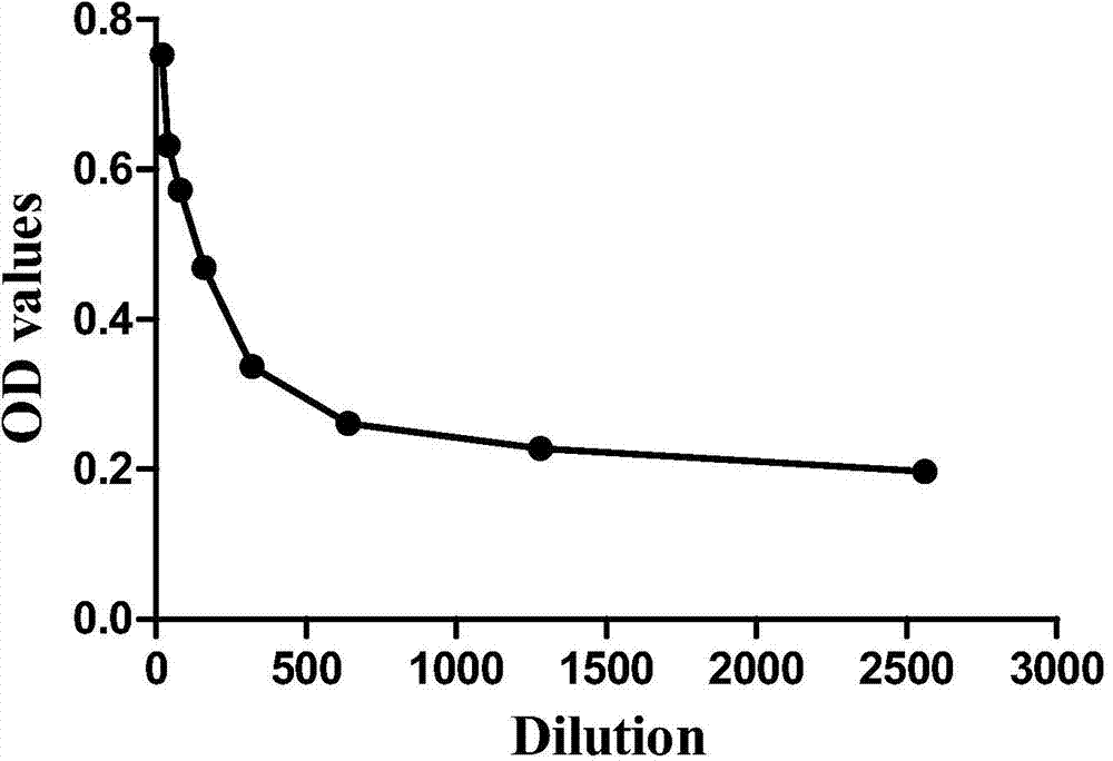
Embodiment 1
[0035] 1) Dissolve and dilute West African simplicifolia agglutinin (Bandeiraea simplicifolia, BS-I) with pH = 9.6 carbonate buffer to 3 μg / mL, add it to each polystyrene reaction well of a 96-well plate, and make It covered the entire bottom of the well, and 100 μL of the lectin solution was added to each well, and left overnight at 4°C.
[0036] 2) The next day, the lectin solution in the wells was discarded, and 200 μL of blocking solution was added to each well to block the parts not bound to the lectin, and incubated at room temperature for 1 h.
[0037] 3) Wash the 96-well plate 4 times with washing buffer. The washing buffer should fill up the entire reaction well each time, shake gently for 3 minutes, discard the liquid in the well and pat dry on absorbent paper.
[0038] 4) Dilute serum samples with PBS (pH=7.4) containing 0.01g / mL BSA at 1:20, 1:40, 1:80, 1:160, 1:320, 1:640, 1:1280, 1: After 2560-fold dilution, add 100 μL to each well of a 96-well plate and incubat...
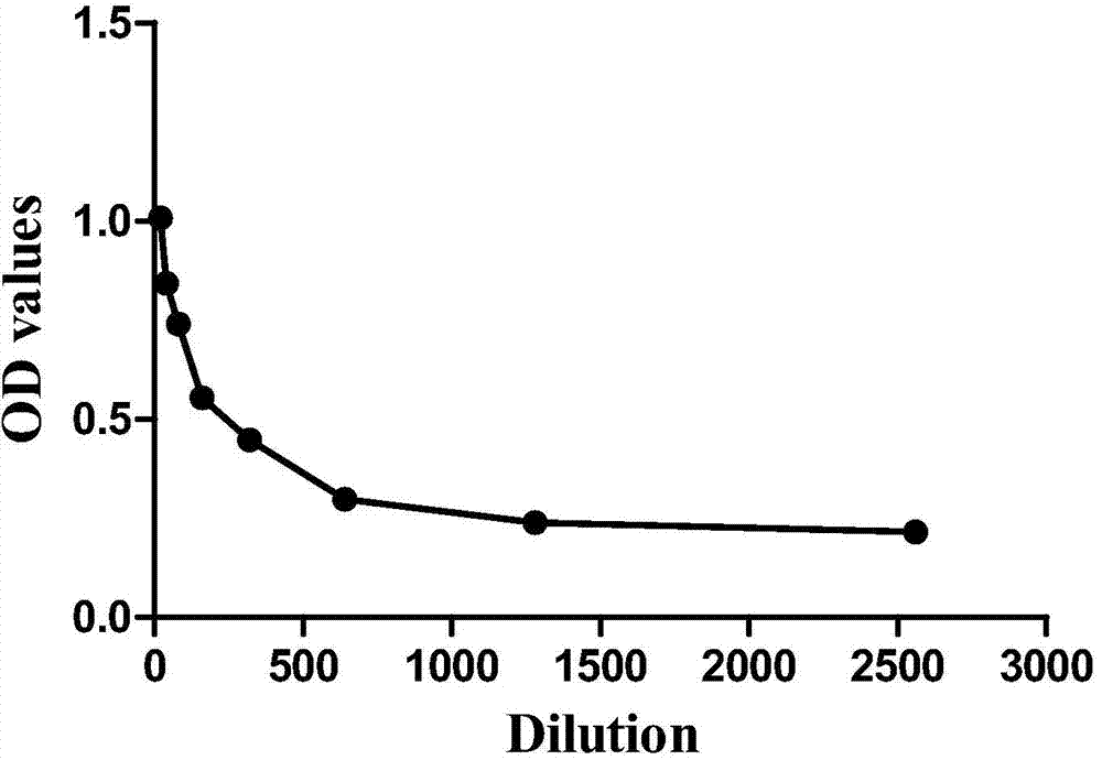
Embodiment 2
[0046] 1) Dissolve Aleuria Aurantia Lectin (AAL) in carbonate buffer solution with pH=9.6 and dilute it to 3 μg / mL, add it into each polystyrene reaction well of a 96-well plate, and make It covered the entire bottom of the well, and 100 μL of the lectin solution was added to each well, and left overnight at 4°C.
[0047] 2) The next day, the lectin solution in the wells was discarded, and 200 μL of blocking solution was added to each well to block the parts not bound to the lectin, and incubated at room temperature for 1 h.
[0048] 3) Wash the 96-well plate 4 times with washing buffer. The washing buffer should fill up the entire reaction well each time, shake gently for 3 minutes, discard the liquid in the well and pat dry on absorbent paper.
[0049] 4) Serum samples were mixed with PBS (pH=7.4) containing 0.01g / mL BSA at 1:20, 1:40, 1:80, 1:160, 1:320, 1:640, 1:1280, 1: 2560-fold dilutions were added to 96-well plates, 100 μL was added to each well, and incubated at room...
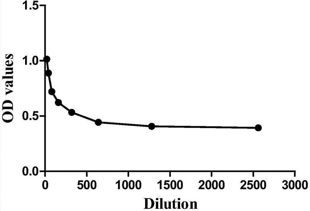
Embodiment 3
[0057] 1) Dissolve and dilute Phytolacca americana (PWM) with pH = 9.6 carbonate buffer to 3 μg / mL, and add it to each polystyrene reaction well of a 96-well plate so that it covers the entire At the bottom of the well, add 100 μL of the lectin solution to each well, and leave overnight at 4°C.
[0058] 2) The next day, the lectin solution in the wells was discarded, and 200 μL of blocking solution was added to each well to block the parts not bound to the lectin, and incubated at room temperature for 1 h.
[0059] 3) Wash the 96-well plate 4 times with washing buffer. The washing buffer should fill up the entire reaction well each time, shake gently for 3 minutes, discard the liquid in the well and pat dry on absorbent paper.
[0060] 4) Serum samples were mixed with PBS (pH=7.4) containing 0.01g / mL BSA at 1:20, 1:40, 1:80, 1:160, 1:320, 1:640, 1:1280, 1: 2560-fold dilutions were added to 96-well plates, 100 μL was added to each well, and incubated at room temperature for 1 ...
PUM
 Abstract
Description
Claims
Application Information