
Fluorescent probe targeting MUC1 for in-vivo ovarian cancer tissue and preparation method of fluorescent probe

A fluorescent probe, ovarian cancer technology, applied in fluorescence/phosphorescence, chemical instruments and methods, luminescent materials, etc., to achieve the effect of increased expression and strong penetration ability

Inactive Publication Date: 2015-04-29
THE SECOND HOSPITAL AFFILIATED TO WENZHOU MEDICAL COLLEGE
View PDF5 Cites 6 Cited by
  • Summary
  • Abstract
  • Description
  • Claims
  • Application Information

AI Technical Summary

Problems solved by technology

At present, various imaging methods used clinically, such as ultrasound, X-ray, CT, MRI, and PET, have been used for tumor detection and preoperative evaluation, but these traditional imaging methods are not suitable for complete high-contrast differentiation and intraoperative real-time identification. There are still some limitations in tumor tissue and normal tissue

Method used


Image

  • Fluorescent probe targeting MUC1 for in-vivo ovarian cancer tissue and preparation method of fluorescent probe
  • Fluorescent probe targeting MUC1 for in-vivo ovarian cancer tissue and preparation method of fluorescent probe
  • Fluorescent probe targeting MUC1 for in-vivo ovarian cancer tissue and preparation method of fluorescent probe

Examples


example 1

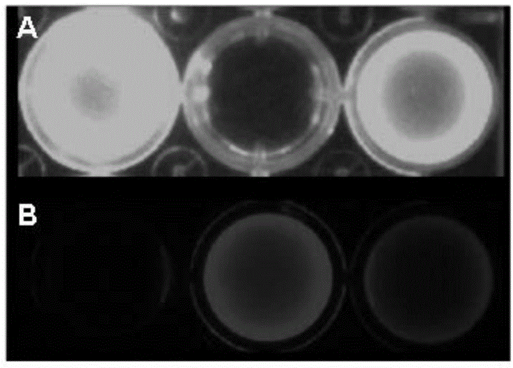
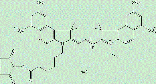
[0022] Example 1: Dilute the anti-MUC1 antibody with 0.1M sodium bicarbonate buffer with a pH value of 8.5 to a final concentration of 1 mg / ml, and then mix the anti-MUC1 antibody / fluorescent dye / Cy5.5 in a ratio of 1:1:1 Labeling was carried out by reacting at 4oC in the dark for 2 hours, and the reactants were filtered and chromatographed through a desalting column (Zeba®, Thermo Scientific, USA) to remove unreacted dyes to obtain the fluorescent probe targeting MUC1 of the living ovarian cancer tissue , by detecting the antibody and bound dye content in a UV spectrophotometer as 1 / 0.7 / 0.8.

example 2

[0023] Example 2: Dilute the anti-MUC1 antibody with 0.1M sodium bicarbonate buffer with a pH value of 8.5 to a final concentration of 1 mg / ml, then anti-MUC1 antibody / fluorescent dye / Cy5.5 at a ratio of 1:10:10 at 4oC After 2 hours of dark reaction for labeling, the reactant was filtered and chromatographed through a desalting column to remove unreacted dye to obtain the fluorescent probe targeting MUC1 of the living ovarian cancer tissue, and detect the antibody and The combined dye content is 1 / 2.5 / 2.8.

example 3

[0024] Example 3: Dilute the anti-MUC1 antibody with 0.1M sodium bicarbonate buffer with a pH value of 8.5 to a final concentration of 1 mg / ml, then anti-MUC1 antibody / fluorescent dye / Cy5.5 at 4oC at a ratio of 1:20:20 After 2 hours of dark reaction for labeling, the reactant was filtered and chromatographed through a desalting column to remove unreacted dye to obtain the fluorescent probe targeting MUC1 of the living ovarian cancer tissue, and detect the antibody and The combined dye content is 1 / 1.9 / 2.0.


PUM


Abstract

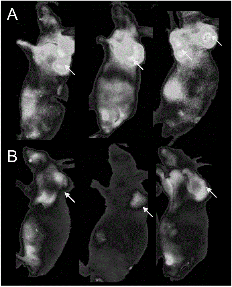
The invention relates to a fluorescent probe and a preparation method thereof in the technical field of biology, particularly to a fluorescent probe targeting MUC1 of in-vivo ovarian cancer tissue, and the preparation method of the fluorescent probe. The fluorescent probe targeting MUC1 of the in-vivo ovarian cancer tissue consists of an antibody against the MUC1, Cy5.5-n-hydroxy succinimide, and a fluorescein NHS dye. Because the fluorescent probe adopts double marks of the near-infrared fluorescent dye cy5.5 and the fluorescein NHS, which are respectively suitable for in-vivo detection and ex-vivo detection; the MUC1 is I type transmembrane glycoprotein which expresses in various kinds of tissue, organ epithelial cells close to lumens, or glandular cavity surfaces, presents top end secretion and is in extreme distribution, and the expressing quantity of the MUC1 in cancer cells is obviously increased. Researches prove that ovarian cancer cells highly express the MUC1, and therefore, the MUC1 can be used as an ideal target for the treatment of ovarian cancer; the fluorescent probe targeting the MUC1, which is combined with double marks, can completely realize the specificity imaging of the ovarian cancer of a living body, so that real-time tracking in operations is completed.

Description

technical field [0001] The invention relates to a fluorescent probe in the field of biotechnology and a preparation method thereof, in particular to a fluorescent probe targeting MUC1 of living ovarian cancer tissue and a preparation method thereof. Background technique [0002] Ovarian cancer is one of the most common malignant tumors in women, and its mortality rate ranks first among gynecological malignancies. Surgery is the mainstay of treatment for ovarian cancer, but the prognosis of patients with advanced ovarian cancer remains poor. During cytoreductive surgery in patients with ovarian cancer, removing as much diseased tissue as possible can improve the efficacy of other treatments. Among them, it is very important to accurately distinguish the boundaries between malignant tumors and surrounding normal tissues or benign lesions. At present, various imaging methods used clinically, such as ultrasound, X-ray, CT, MRI, and PET, have been used for tumor detection and p...

Claims


Application Information

Patent Timeline
IPC IPC(8): C09K11/06G01N21/64
Inventor 张琼朱雪琼武垚森沈奇卢晓声林焱
Owner THE SECOND HOSPITAL AFFILIATED TO WENZHOU MEDICAL COLLEGE
Learn More