Living lung slicing based anti-influenza virus or anti-inflammatory drug screening model establishing method and application

A technology of anti-influenza virus and anti-inflammatory drugs, applied in the field of medicine, can solve the problems of inaccurate evaluation, no drug screening, no development of drug evaluation models, etc., and achieve the effect of saving experimental materials and saving usage

Inactive Publication Date: 2015-09-02
WUHAN INST OF VIROLOGY CHINESE ACADEMY OF SCI
View PDF0 Cites 3 Cited by
  • Summary
  • Abstract
  • Description
  • Claims
  • Application Information

AI Technical Summary

Problems solved by technology

However, in the cell model, because the isolated single cell loses its tissue environment, it cannot simulate its relevant functions in the body well, and the evaluation is not accurate.
Drugs that work on cells often don't work as well in animals
Although biopsies

Method used


Image

  • Living lung slicing based anti-influenza virus or anti-inflammatory drug screening model establishing method and application
  • Living lung slicing based anti-influenza virus or anti-inflammatory drug screening model establishing method and application
  • Living lung slicing based anti-influenza virus or anti-inflammatory drug screening model establishing method and application

Examples


Embodiment 1

[0058] Example 1 Preparation of lung slices

[0059] (1) After the mice were anesthetized by intraperitoneal injection of pentobarbital (75 mg / kg), they were sacrificed by exsanguination from the abdominal aorta. The abdominal skin was wiped and disinfected with alcohol cotton balls. After the trachea was exposed, the trachea was separated with ophthalmic forceps, and a "V"-shaped incision was made at the trachea (centrifugal end) with ophthalmic scissors. Insert an 18-gauge ground syringe needle through the "V" incision and secure with suture. Then fill the lungs with 1.3ml of low-melting gel preheated to 37°C.

[0060] (2) Place the whole mouse on ice for 10 minutes to allow the gel to initially solidify. After the gel solidified, cut off the trachea, carefully remove the heart and the whole lung, put them in pre-cooled DMEM / F12 medium, and place them at 4°C for 15 minutes to make the gel completely solidify. Separate the lung and select the left lung lobe. Use ophthalm...

Embodiment 2

[0063] Embodiment 2 The thickness of lung section is preferred

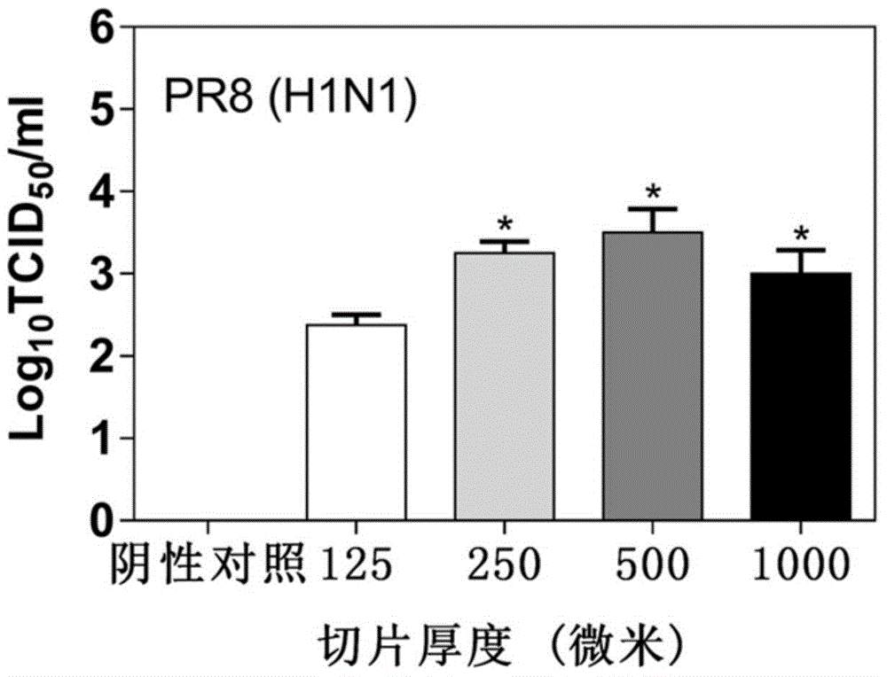
[0064] (1) with 10 5 H1N1 virus infection of PFU / ml Lung slice sample 1, sample 2, sample 3 and sample 4 prepared in Example 1. After 2 hours of infection, the virus solution was discarded, washed twice with phosphate buffer, and fresh culture solution was added.

[0065] (2) Collect the supernatant after 48 hours. Tissue median infectious dose (TCID) 50 ) to detect the virus titer in the section supernatant, and the virus titer was calculated using the Reed–Muench method (Reed, L.J., Muench, H., 1938. American Journal of Epidemiology 27, 493-497.). The result is as figure 1 As shown, it can be seen that the log in samples 1-4 10 The TCID50 values ​​were 2.375, 3.25, 3.5, and 3, respectively. The virus particles in the supernatant of sample 2 were 7.5 times more than those in the lung slice supernatant of sample 1, but the difference was not obvious from sample 3 and sample 4. In sample 2-sample 4, the thic...

Embodiment 3

[0066] Example 3 Neuraminidase (NA) activity and viral activity correlation evaluation

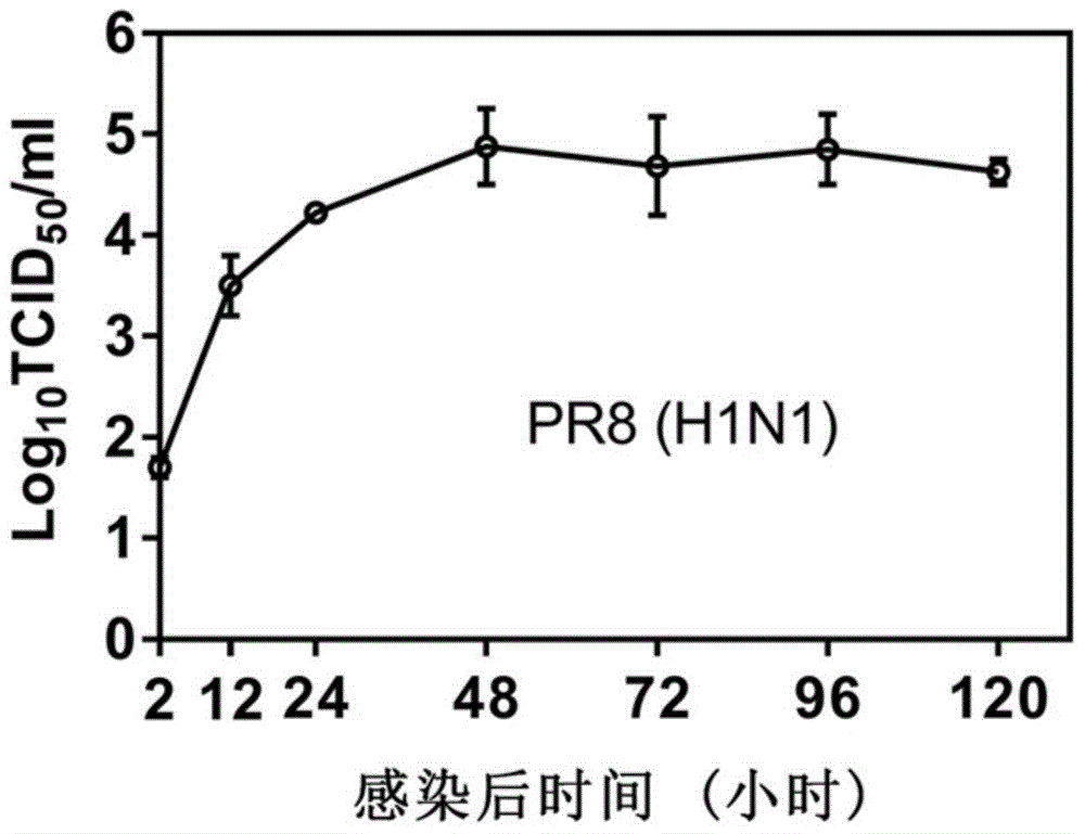
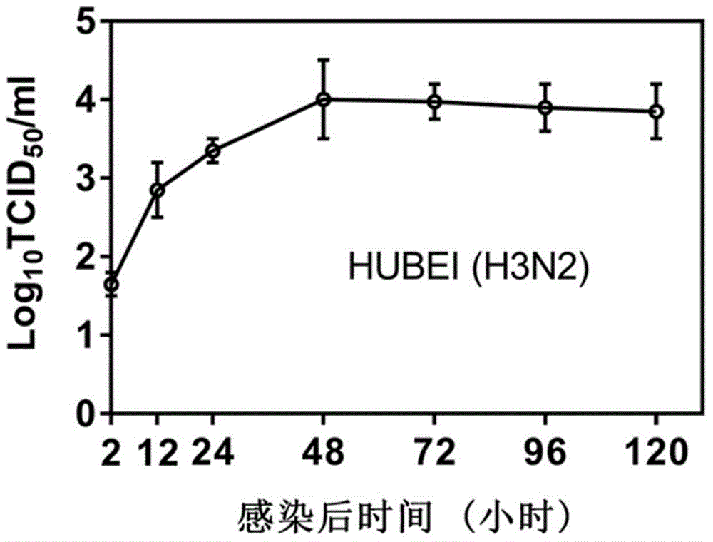
[0067] (1) with 10 5 PFU / ml H1N1 virus or H3N2 virus infection lung slice sample 2 prepared in Example 1. After 2 hours of infection, the virus solution was discarded, washed twice with phosphate buffer, and fresh culture solution was added.

[0068] (2) The supernatants of lung slice samples were collected after 12 hours, 24 hours, 48 ​​hours, 72 hours, 96 hours and 120 hours.

[0069] (3) The virus titer in the supernatant collected in step (2) was detected by tissue half infectious dose (TCID50), and the virus titer was calculated by the Reed-Muench method. The result is as Figure 2a , 2b As shown, the virus reached the replication peak at 48h and entered the stable replication stage.

[0070] (4) 2'-(4-methylumbelliferone)-α-D-acetylneuraminic acid (MUNANA) is a specific fluorescent substrate for neuraminidase (NA), used to express neuraminidase active. Put 40 μl of the virus s...


PUM

PropertyMeasurementUnit
Thicknessaaaaaaaaaa
Thicknessaaaaaaaaaa

Abstract

The invention discloses a living lung slicing based anti-influenza virus or anti-inflammatory drug screening model establishing method and application. The method is characterized by including the following steps of firstly, slicing the virus-infected living lung; secondly, allowing to-be-detected drugs to act on slices of the virus-infected living lung; finally, verifying effect of the to-be-detected drugs by detecting changes of virus titer and related cytokines. The invention further discloses a screening model establishing method which includes: preparation of the lung slices, optimal thickness selection of the lung slices, relevant evaluation of neuraminidase and the virus titer and selection of inflammatory factors. The drug screening method on the basis of living lung slicing is established, and compared with a traditional cell model, the living lung slicing based anti-influenza virus or anti-inflammatory drug screening model establishing method is more in line with true body conditions and is more effective in accuracy.

Description

technical field [0001] The invention belongs to the technical field of medicine, and more specifically relates to a construction method and application of an anti-influenza virus or anti-inflammatory drug screening model based on living lung slices. Background technique [0002] Influenza virus infection seriously threatens human health and brings serious economic losses to society. The 1918 influenza pandemic infected about 500 million people worldwide, resulting in 50-100 million deaths. The swine flu outbreak in 2009 spread to more than 200 countries and killed about 18,000 people. In 2013, the emergence of highly pathogenic avian influenza H7N9 in China once again aroused people's attention on influenza viruses and how to control influenza. In addition to influenza outbreaks, annual seasonal influenza also brings great losses to society, with approximately 250,000 to 500,000 deaths from influenza worldwide. Currently, the main treatment strategy for influenza is antiv...

Claims


Application Information

Patent Timeline
IPC IPC(8): C12Q1/44G01N33/68
Inventor 陈绪林刘锐
Owner WUHAN INST OF VIROLOGY CHINESE ACADEMY OF SCI
Why Eureka
Try Eureka