External fixation support formed through 3D printing and used for personalized orthopedic and manufacturing method thereof

A 3D printing and external fixation technology, applied in medical science and other directions, can solve problems such as breathing difficulties, achieve good air permeability, avoid complications, and ensure the effect of air permeability

Inactive Publication Date: 2015-09-16
JIANGYIN RECONGENE BIOMEDICAL TECH
View PDF5 Cites 27 Cited by
  • Summary
  • Abstract
  • Description
  • Claims
  • Application Information

AI Technical Summary

Problems solved by technology

In addition, after the plaster vest is fixed on the chest and upper abdomen, if the fixation is too tight, the expansion of the stomach and thorax will be restricted, resulting in symptoms such as chest tightness, dyspnea, abdominal pain, and vomiting

Method used


Image

  • External fixation support formed through 3D printing and used for personalized orthopedic and manufacturing method thereof

Examples


Embodiment 1

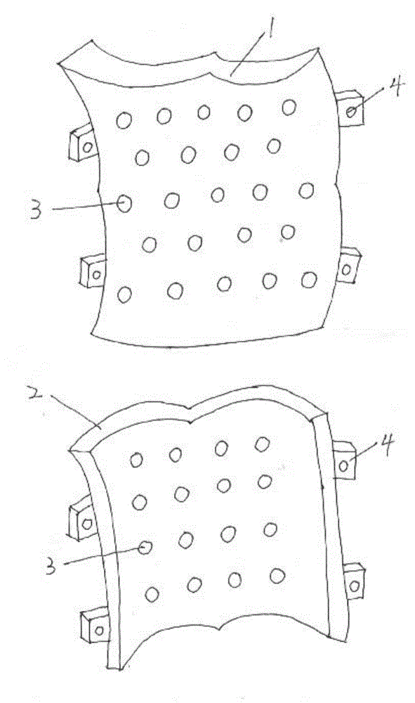
[0029] Embodiment 1 A method of utilizing 3D printing to prepare a personalized orthopedic external fixation bracket, the specific steps are as follows:

[0030] 1) Use a photographic 3D scanner to perform 3D scanning on the part of the patient that needs external fixation, and obtain the surface 3D point data of the part; at this time, the patient needs to perform 3D scanning in an upright suspension traction state under the guidance of the doctor after the operation ;

[0031] 2) Process the acquired 3D data with the 3D reverse engineering software mimics10.01 (MATERIALISE, Belgium) to generate a 3D model of the patient’s body parts that require external fixation; The 3D digital model of the scanned part lays the foundation for the next step of stent design;

[0032] 3) Based on the 3D model of the above-mentioned parts of the human body that require external fixation, use the software magics (MATERIALISE company, Belgium) and software solidworks (Solidworks, the United Sta...


PUM


Abstract

The invention discloses a method for manufacturing an external fixation support for personalized orthopedic by utilizing 3D printing. The method comprises the steps of conducting three-dimensional scanning on a postoperative patient in a traction state to obtain outer surface three-dimensional data of the position needed to be externally fixed of the postoperative patient, re-establishing a three-dimensional model of the position needed to be externally fixed according to the three-dimensional data, using software to design an external fixation support completely fit with the position needed to be externally of the patient and used for orthopedic, arranging air-permeable holes and fixing blocks on the support model and using polylactic acid as a raw material to obtain the external fixation support for orthopedic through 3D printing performed by a 3D printer. By means of the method, the external fixation support completely fit with the position needed to be externally of the patient and used for orthopedic can be manufactured, long-time shaping through plaster fixation is avoided, the degree of tightness can be adjusted, the external fixation support is light and air-permeable, and a more comfortable fixation mode is provided for the patient.

Description

technical field [0001] The invention relates to a method for preparing an external fixation bracket for orthopedics, in particular to a method for preparing a personalized external fixation bracket for orthopedics by 3D printing and the external fixation bracket printed by the method. Background technique [0002] Since Mager first reported its application in clinical treatment of thoracolumbar fractures in 1982, it has been welcomed by clinicians because of its advantages of less trauma and less bleeding. The industry generally believes that when the internal fixation is not enough to maintain the stability of the spine, it is an effective method to protect the spine with external fixation until the bone graft heals firmly. Especially after spinal instability and total spinal osteotomy, reliable external fixation is required to avoid postoperative accidents and fusion failure. [0003] Gypsum is easy to obtain, cheap, has good plasticity and sufficient strength, and is cur...

Claims


Application Information

Patent Timeline
Patent Type & Authority Applications(China)
IPC IPC(8): A61F5/01
Inventor 史建刚林腾飞刘青孟潇
Owner JIANGYIN RECONGENE BIOMEDICAL TECH
Why Eureka
Try Eureka