Coronary three-dimensional reconstruction calcified plaque removing method

A calcified plaque and three-dimensional reconstruction technology, applied in the field of calcified plaque removal, can solve the problems of inaccurate coronary three-dimensional reconstruction, wrong stenosis estimation, stenosis, etc.

Inactive Publication Date: 2015-11-25
北京欣方悦医疗科技有限公司
View PDF5 Cites 12 Cited by
  • Summary
  • Abstract
  • Description
  • Claims
  • Application Information

AI Technical Summary

Problems solved by technology

This will result in inaccuracy of coronary 3D reconstruction
In particular, calcified plaque usually causes stenosis, and blood vessels that have not comple...

Method used


Image

  • Coronary three-dimensional reconstruction calcified plaque removing method
  • Coronary three-dimensional reconstruction calcified plaque removing method
  • Coronary three-dimensional reconstruction calcified plaque removing method

Examples


Embodiment Construction

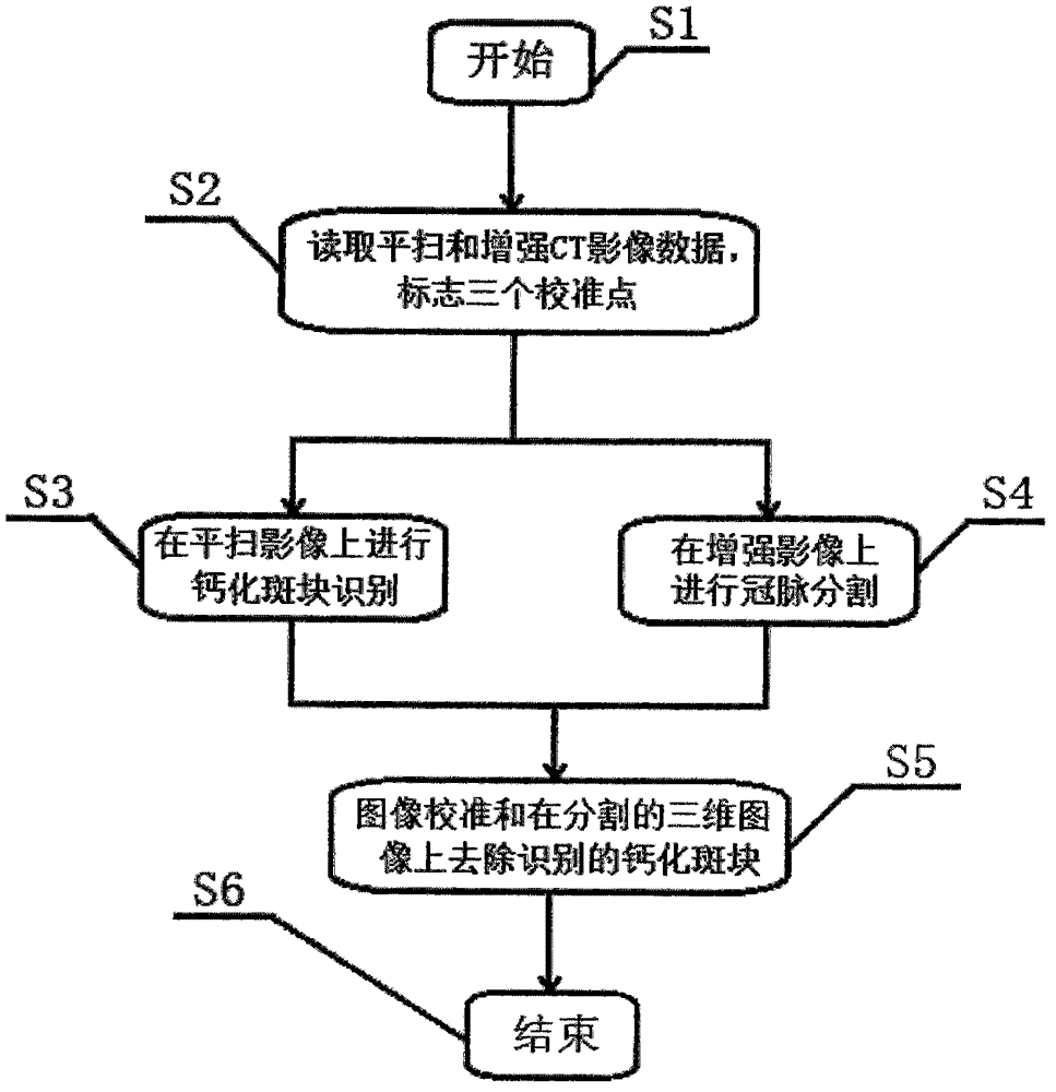
[0040] Such as figure 1 Shown, concrete steps of the present invention are:

[0041] In step S1, start and enter step S2;

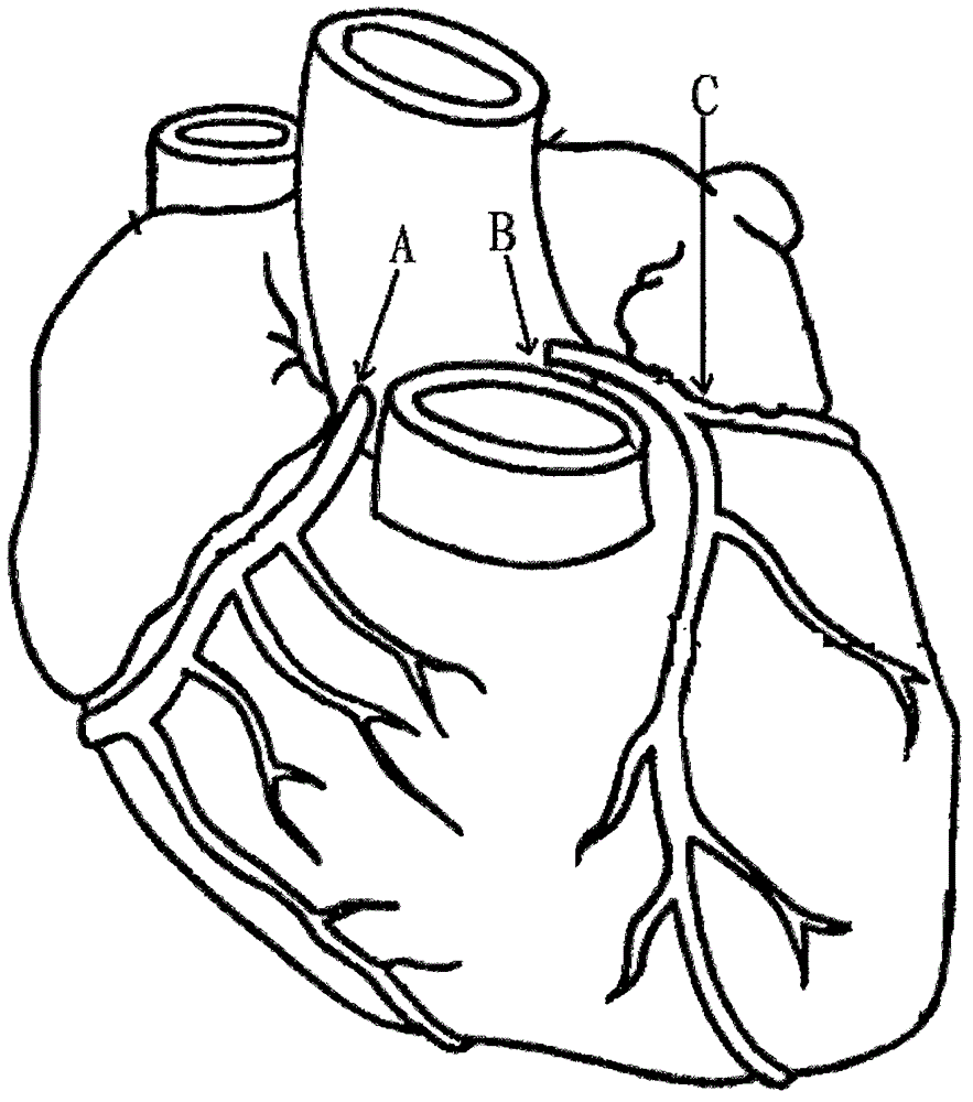
[0042] In step S2, the plain scan CT and enhanced CT image data of the patient are read; in a specific CT scan, the two scans can be completed at the same time, or in different time periods. If divided into time periods, the body positions and scanning angles of the two scans are required to be consistent. Then select three calibration points A, B, and C respectively on the image data. Calibration points are used to calibrate two different image data into the same coordinate space. Calibration points can be selected from the entrance of the left crown, the entrance of the right crown and the first bifurcation point of the left crown (the bifurcation point of the left anterior descending branch and the left circumflex branch) or other bifurcation points, see figure 2 Points A, B and C marked by arrows. Of course, other points can also be selected for...


PUM


Abstract

The invention discloses a coronary three-dimensional reconstruction calcified plaque removing method. The concrete steps of the method are divided into six steps: in the step S1, starting is performed; in the step S2, non-enhanced CT and enhanced CT image data of a patient are read; in the step S3, calcified plaque identification is performed on the non-enhanced image data; in the step S4, coronary segmentation and three-dimensional coronary model reconstruction are performed on the enhanced CT image; in the step S5, two three-dimensional spaces of the calcified plaque and a coronary grid are calibrated and the calcified plaque and the coronary grid are arranged in the same coordinate system; finally the obtained grid is a coronary blood three-dimensional image model of which the calcified plaque is removed; and the process enters the step S6 and ends. The non-enhanced CT and the enhanced CT are compared so that the non-enhanced identification calcified plaque can be removed from the enhanced CT coronary three-dimensional model, and an accurate coronary three-dimensional blood form model can be acquired; and support is provided for clinic narrow assessment and hydrodynamic blood dynamic analysis.

Description

technical field [0001] The invention relates to a method for removing calcified plaque, in particular to a method for removing calcified plaque in coronary three-dimensional reconstruction. Background technique [0002] The technology closest to the present invention is the blood vessel image segmentation technology based on medical imaging. Its principle is to use a growth algorithm to segment blood vessels according to the gray scale range of pixels, so as to obtain a three-dimensional image model. In order to perform better image segmentation, this type of algorithm is mainly based on enhanced CT (contrast agent injected intravenously by the patient before scanning to increase the brightness of blood under X-ray), according to the enhanced brightness of the blood gray value range from the surrounding tissue, using Region growing algorithm for coronary artery segmentation and 3D image reconstruction. [0003] Existing techniques cannot deal with calcified plaque well. Ac...

Claims


Application Information

Patent Timeline
IPC IPC(8): G06T5/00G06T17/00A61B6/03
Inventor 唐铁兵
Owner 北京欣方悦医疗科技有限公司
Why Eureka
Try Eureka