A Fast and Robust Automatic Segmentation Method for Liver in Abdominal CT Sequence Images

A sequential image and automatic segmentation technology, applied in the field of image processing, can solve the problems of long time, poor segmentation effect, sensitive data initialization and registration, etc., and achieve the effect of suppressing complex background, fast segmentation and strong robustness.

Active Publication Date: 2017-09-05
湖南提奥医疗科技有限公司
View PDF3 Cites 2 Cited by
  • Summary
  • Abstract
  • Description
  • Claims
  • Application Information

AI Technical Summary

Problems solved by technology

The purely image-based segmentation method refers to the method of directly using brightness, texture and other image information for segmentation, mainly including threshold method, clustering, region growing, active contour model and graph cut, etc. These segmentation techniques mainly have the following disadvantages and Disadvantages: (1) Complex preprocessing is required, including removal of surrounding tissues or organs such as ribs, spine, and kidneys; (2) It is difficult to segment CT sequence images with low contrast and blurred liver boundaries
The method based on the statistical model first uses a large number of CT sequence images to construct the target prior model, and then applies it to the segmentation of the current sequence. This type of method has a better segmentation effect for images with low contrast, but for irregular liver The segmentation effect is poor, it takes a long time, and it is sensitive to data initialization and registration

Method used


Image

  • A Fast and Robust Automatic Segmentation Method for Liver in Abdominal CT Sequence Images
  • A Fast and Robust Automatic Segmentation Method for Liver in Abdominal CT Sequence Images
  • A Fast and Robust Automatic Segmentation Method for Liver in Abdominal CT Sequence Images

Examples


Embodiment 1

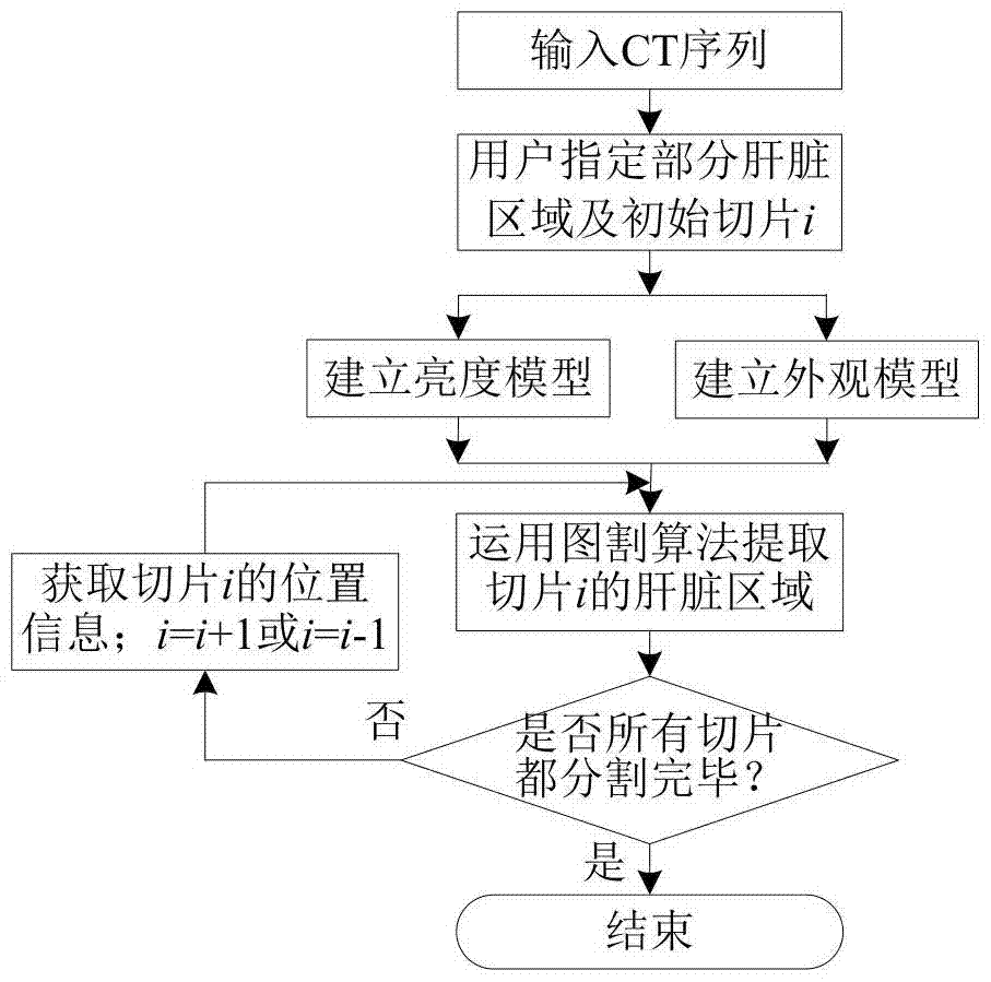
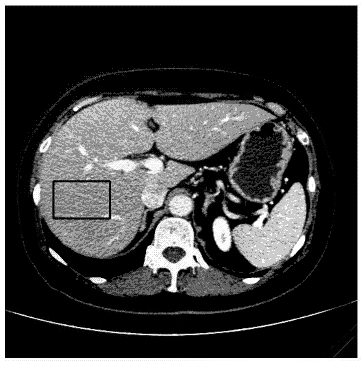
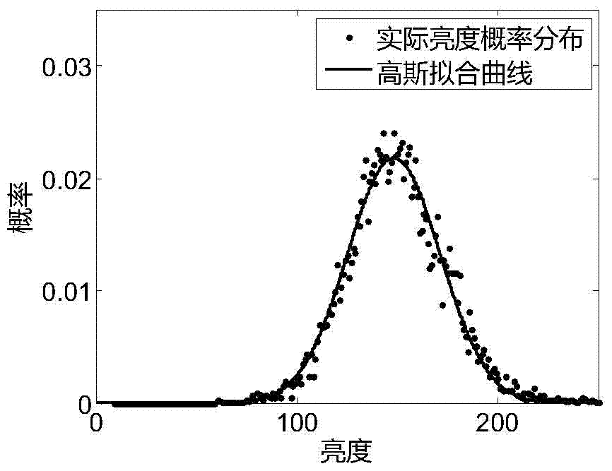
[0044] figure 1Shown is a flowchart of a method for robust automatic liver segmentation of abdominal CT sequence images according to an embodiment of the present invention. First, a part of the liver area is arbitrarily selected from the input CT sequence to establish a liver brightness model and an appearance model, and then the liver in the initial slice is segmented using the graph cut algorithm combined with the brightness and appearance model, and finally the initial segmentation slice is iteratively The starting point splits the liver of all other slices in the sequence up and down, respectively. In the iterative segmentation process, the liver position information of the previous segmentation result is integrated into the graph cut energy function of the current slice to increase the accuracy of the segmentation result. Until all slices are segmented, the program runs to the end, and the segmented results are output.

[0045] Combine below figure 1 , using a preferre...

Embodiment 2

[0080] The method of Example 1 is used to test 10 liver CT sequences provided by the XHCSU14 database, and five error indicators are used to evaluate the test results, including: volume overlap error (Volumetric Overlap Error, VOE), relative volume difference (RelativeVolumeDifference, RVD), average symmetrical surface distance (Average SymmetricSurfaceDistance, ASD), root mean square symmetrical surface distance (Root Mean Square SymmetricSurfaceDistance, RMSD), and maximum symmetrical surface distance (Maximum Symmetric SurfaceDistance, MSD).

[0081] The 10 test sequences of the XHCSU14 database are all derived from the Philipsbrilliance 64-slice multi-slice spiral CT machine provided by Xiangya Hospital of Central South University. mm. The segmentation results of the XHCSU14 database were evaluated using five error indicators: VOE, RVD, ASD, RMSD, and MSD. The results are shown in Table 1. It can be seen that for the CT sequences of 10 different patients, the mean value a...


PUM


Abstract

The invention discloses a robustness auto-partitioning method for an abdomen computed tomography (CT) sequence image of a liver. The robustness auto-partitioning method comprises a data inputting step : in which a CT sequence to be partitioned is input and an initial slice is designated; a model building step in which a liver brightness model and an appearance model are built according to data characteristics of the input sequence,a complex background is suppressed and a liver region is highlighted; and an automatic partitioning step in which the initial slice is rapidly and automatically partitioned through combining the brightness model and the appearance model by a graph cut algorithm, and all slices in the liver CT sequence are iteratively partitioned upwards and downwards by taking the initial partition slice as a starting point according to spatial correlations between adjacent slices. According to the method, the corresponding brightness and appearance models are built with regards to the particular CT sequence, and thus, the liver with a low partitioning contrast ratio, boundary fuzziness and shape irregularity can be effectively and automatically partitioned. Moreover, the auto-partitioning method for the abdomen CT sequence image of the liver can be promoted to automatic partitioning of other abdominal organs, such as partitioning of the abdomen CT sequence image of a spleen and a kidney.

Description

technical field [0001] The invention belongs to the technical field of image processing, and relates to the segmentation of organs in abdominal CT sequence images, in particular to the fast and robust automatic segmentation of liver in abdominal CT sequence images, which can be used for medical image auxiliary diagnosis and treatment. Background technique [0002] Liver segmentation is a prerequisite for computer-aided diagnosis of liver diseases and preoperative planning for liver transplantation. The liver model obtained by segmentation and reconstruction can assist in liver lesion analysis, volume measurement, blood vessel analysis, liver segmentation, disease diagnosis and evaluation, etc. CT angiography images are generally favored by doctors because of their high resolution, little damage to the human body, and their ability to vividly and accurately reflect the location of the liver and its lesions. Due to the large number of image slices used in 3D imaging (about 20...

Claims


Application Information

Patent Timeline
Patent Type & Authority Patents(China)
IPC IPC(8): G06T7/11G06T7/187
CPCG06T2207/10081G06T2207/30056
Inventor 赵于前廖苗
Owner 湖南提奥医疗科技有限公司
Why Eureka
Try Eureka