
Phased array head temporal bone imager

A phased array, imager technology, applied in ultrasonic/sonic/infrasound image/data processing, ultrasonic/sonic/infrasonic Permian technology, organ motion/change detection, etc., can solve problems such as high inspection costs, Achieve the effect of simple structure, small appearance and low inspection cost

Inactive Publication Date: 2015-12-16
苏州斯科特医学影像科技有限公司
View PDF12 Cites 4 Cited by
  • Summary
  • Abstract
  • Description
  • Claims
  • Application Information

AI Technical Summary

Problems solved by technology

The above examinations have X-ray radiation on the patient, and the examination methods must be examined in the radiology department, CT room, and MR large-scale equipment room of medical imaging, and the examination fee is also very expensive.
There is currently no dedicated device for brain diagnostics using ultrasound

Method used


Image

  • Phased array head temporal bone imager

Examples


Embodiment Construction

[0012] The present invention will be further described below in conjunction with accompanying drawing.

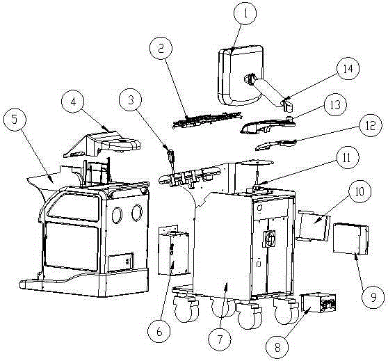
[0013] Phased array craniotemporal bone imager, including display main body 1, main input device 2, ultrasonic probe 3, auxiliary input device 4, shell 5, ultrasonic host 6, body 7, power supply 8, PC host 9, optical drive 10, display guide column 11. Display bottom cover 12, display arm 13, display bracket 14, characterized in that the display body 1 is set on the display arm 13 through the display bracket 14, the display arm 13 is provided with the display bottom cover 12, and the display guide column 11 passes through the display Bottom cover 12, display arm 13 and display bottom cover 12, display arm 13 are connected on the body 7, PC host 9, optical drive 10 are arranged on the rear side of body 7, power supply 8 is arranged on the bottom of PC host 9, optical drive 10, ultrasonic The host 6 is arranged in front of the body 7, and the body 7 is wrapped by the shell 5. ...


PUM


Abstract

The invention relates to a phased array head temporal bone imager. The phased array head temporal bone imager comprises a displayer body, a main input device, an ultrasonic probe, an auxiliary input device, an outer shell, an ultrasonic main machine, a machine body, a power source, a PC host, a CD driver, a displayer guide column, a displayer bottom cover, a displayer arm and a displayer support. When the phased array head temporal bone imager is in practical use, the 2-MHz low-frequency phased array probe is adopted to penetrate through the head temporal bone to observe a brain tissue structure, intracranial vessels and vessel conditions of a wemes ring of the brainstem, and intracranial lesions are diagnosed through the brain parenchyma, the brain vessels and Doppler frequency spectrum waveforms of the brain parenchyma and the brain vessels. When used for detection, the phased array head temporal bone imager is direct, easy, convenient, free of radiation and low in detection cost. Meanwhile, blood flowing and pulsation can be observed in real time, and analysis and diagnosis are carried out in cooperation with the conditions such as the rigidity of the vessels and the blood concentration.

Description

technical field [0001] The invention relates to ultrasonic diagnosis and treatment equipment, in particular to a phased array cranial temporal bone imager. Background technique [0002] Ultrasound diagnosis mainly uses the good directivity of ultrasound and physical characteristics such as reflection, scattering, attenuation and Doppler effect similar to light, using different physical parameters, using different types of ultrasonic diagnostic instruments, and using various scanning methods , the ultrasound is transmitted into the human body and propagates in the tissue. When there is a certain difference in the acoustic impedance of normal tissue or pathological tissue, the interface formed by them will reflect and scatter, and then receive the echo signal and perform detection and other processing. After that, it will be displayed as waveform, curve or image, etc. [0003] As a kind of ultrasonic diagnosis, B-ultrasound can clearly display various cross-sectional images o...

Claims


Application Information

Patent Timeline
Patent Type & Authority Applications(China)
IPC IPC(8): A61B8/08
Inventor 高兴斌周正帮吴丽丽
Owner 苏州斯科特医学影像科技有限公司
Learn More