Single-scanning quantitative magnetic resonance diffusion imaging method based on dual echoes

A technology of magnetic resonance imaging and imaging methods, which is applied in the directions of diagnostic recording/measurement, medical science, sensors, etc., can solve the problems of increasing time and signal attenuation, the effect is not good enough, and the imaging efficiency is not greatly improved.

Active Publication Date: 2016-01-13
XIAMEN UNIV
View PDF6 Cites 24 Cited by
  • Summary
  • Abstract
  • Description
  • Claims
  • Application Information

AI Technical Summary

Problems solved by technology

However, this method has limitations. On the one hand, this method needs to extend the echo chain, which will inevitably lead to increased acquisition time and signal attenuation; on the other hand, compared with the conventional EPI method, this method is achieved by extending the repetition time. At the expense of (TR), this may require sacrificing the spatial resolution of the re

Method used


Image

  • Single-scanning quantitative magnetic resonance diffusion imaging method based on dual echoes
  • Single-scanning quantitative magnetic resonance diffusion imaging method based on dual echoes
  • Single-scanning quantitative magnetic resonance diffusion imaging method based on dual echoes

Examples


specific Embodiment

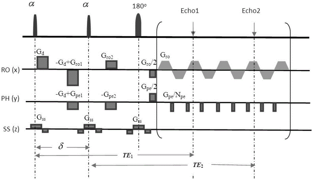
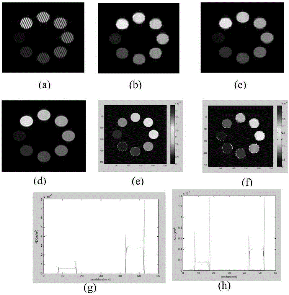
[0060] The feasibility of the present invention is verified by a simulation model experiment based on a double-echo single-scan quantitative magnetic resonance diffusion imaging (DM-OLED) method. Before the experiment, use matlab to generate eight circle models. The model includes ADC, T2, T1, and proton density model. The ADC and T2 values ​​​​of the eight circles are different, ADC=0.67e-9~4.4e-9s / m 2 , T2=0.08~0.1s, T1=1s, this group of T1 and T2 is equivalent to the ratio of T1 and T2 of human tissue under the magnetic field of 3T. Use the ADC model as a reference.

[0061] Simulation steps:

[0062] 1. Set the size of the imaging field (FOV), the imaging field of view FOV in the x direction x 60mm, the imaging field of view FOV in the y direction y is 60mm. Add model files to the appeal;

[0063] 2. Calculate the sequence parameters. The test parameters of the present embodiment are set as follows: the excitation time of the 45° excitation pulse is 3 milliseconds, an...


PUM


Abstract

The invention provides a single-scanning quantitative magnetic resonance diffusion imaging method based on dual echo, and relates to a magnetic resonance imaging method. According to the method, two echoes with the same evolution time are generated through two small-angle excitation pulses with the same turning angle, so that the same transverse relaxation time is achieved; a displacement gradient is added after each excitation pulse to achieve central displacement of the two echo signals in a signal space, and a diffusion gradient is added after the first excitation pulse, so that diffusion reduction only exists in the first echo signal; accordingly, signals under different diffusion factors are obtained. The two echo signals are from one imaging slice, so that the two echo signals can be separated through priori knowledge of the two echo signals by matching sparse conversion with a corresponding separation algorithm. Finally, a quantitative ADC image is obtained by performing apparent diffusion coefficient calculation on two signals obtained through separation. Single-scanning quantitative ADC imaging is obtained through the method, and the quality of the obtained ADC image is good.

Description

technical field [0001] The invention relates to a magnetic resonance imaging method, in particular to a single-scan quantitative magnetic resonance apparent diffusion coefficient (apparent diffusion coefficient, ADC) imaging method. Background technique [0002] Diffusion imaging, as a kind of MRI quantitative imaging, provides a contrast mechanism for analyzing normal and pathological living tissues in a non-invasive way, and has been applied to the study of the microstructure of the brain, heart, and spinal cord. Single-scan ultrafast magnetic resonance imaging (MRI) is of great importance in in vivo diffusion studies. This requires overcoming spontaneous motion in the magnet to measure displacements on the order of arbitrary microseconds in experiments without external disturbances. However, diffusion imaging needs to obtain a series of contrast-weighted images during its imaging process, and the apparent diffusion coefficient (apparent diffusion coefficient, ADC). Exi...

Claims


Application Information

Patent Timeline
IPC IPC(8): A61B5/055
Inventor 蔡聪波马崚嶒陈忠蔡淑惠丁兴号
Owner XIAMEN UNIV
Why Eureka
Try Eureka