Image scattering correction method, device and apparatus

A scattering correction and image technology, applied in the field of medical image processing, can solve problems such as difficult CBCT projection map correction, and achieve effective correction effect

Active Publication Date: 2016-05-11
NEUSOFT MEDICAL SYST CO LTD
View PDF8 Cites 19 Cited by
  • Summary
  • Abstract
  • Description
  • Claims
  • Application Information

AI Technical Summary

Problems solved by technology

[0004] The present invention provides an image scattering correction method, device and equipment to solve the existing problem that it is difficult to effectively correct CBCT projection images

Method used


Image

  • Image scattering correction method, device and apparatus
  • Image scattering correction method, device and apparatus
  • Image scattering correction method, device and apparatus

Examples


Embodiment Construction

[0026] The terminology used in the present invention is for the purpose of describing particular embodiments only and is not intended to limit the invention. As used herein and in the appended claims, the singular forms "a", "the", and "the" are intended to include the plural forms as well, unless the context clearly dictates otherwise. It should also be understood that the term "and / or" as used herein refers to and includes any and all possible combinations of one or more of the associated listed items.

[0027] It should be understood that although the terms first, second, third, etc. may be used in the present invention to describe various information, the information should not be limited to these terms. These terms are only used to distinguish information of the same type from one another. For example, without departing from the scope of the present invention, first information may also be called second information, and similarly, second information may also be called fi...


PUM


Abstract

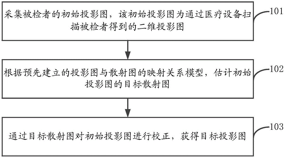
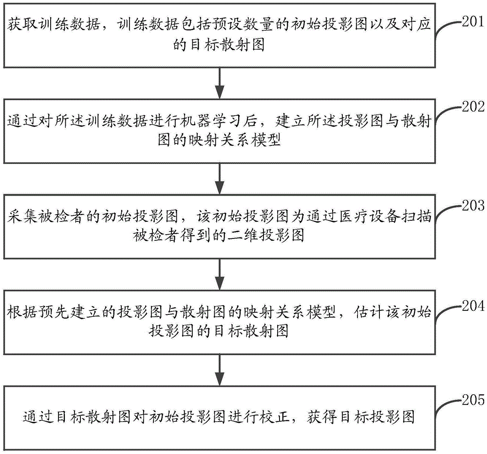
The present invention discloses an image scattering correction method, a device and an apparatus. The method comprises the steps of acquiring the initial projection drawing of a to-be-checked person, wherein the initial projection drawing is in the form of a two-dimensional projection drawing obtained through scanning the to-be-checked person by means of a medical facility; based on a mapping relationship model between the pre-established projection drawing and a scattering diagram, estimating the target scattering diagram of the initial projection drawing; and correcting the initial projection drawing through the target scattering diagram to obtain a target projection drawing. According to the embodiments of the invention, after the initial projection drawing of the to-be-checked person is acquired through scanning the to-be-checked person by the medical facility, the accurate target scattering diagram of the initial projection drawing is estimated based on the mapping relationship model capable of accurately representing the relationship between the pre-established projection drawing and the scattering diagram. After that, the initial projection drawing is corrected through the target scattering diagram. In this way, the accurate target projection drawing can be obtained, so that the initial projection drawing is effectively corrected.

Description

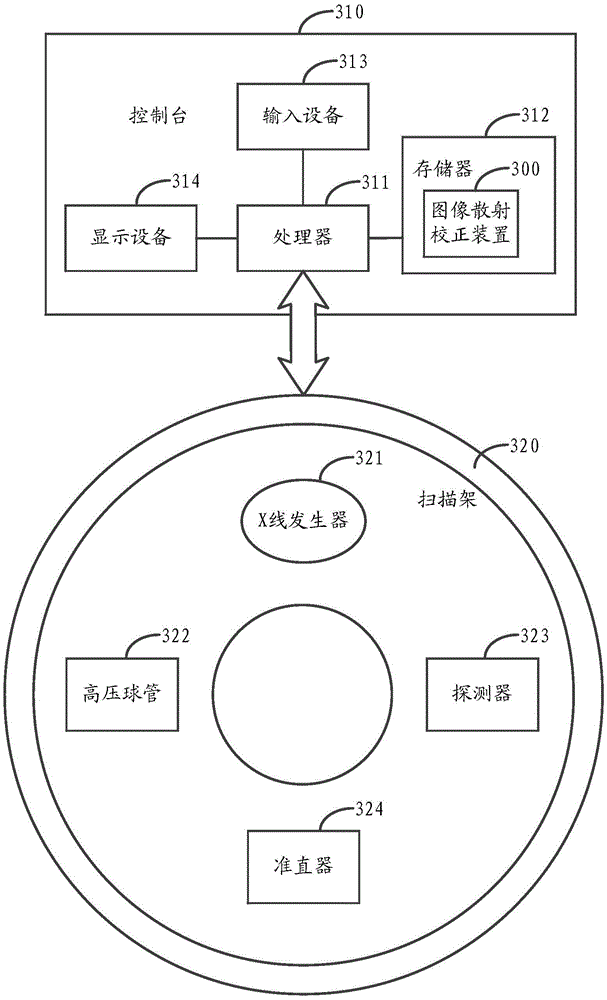
technical field [0001] The invention relates to the technical field of medical image processing, in particular to an image scattering correction method, device and equipment. Background technique [0002] CBCT (Cone Beam Computed Tomography, cone beam X-ray computerized tomography) equipment is a medical imaging equipment widely used in the medical field. When the CBCT equipment is used to scan the subject, the X-ray is emitted by the point source. After the X-ray passes through the subject, it is received by the detection point on the flat-panel detector. According to the number of photons of the X-ray received by the flat-panel detector A two-dimensional CBCT projection map can be generated, and finally a three-dimensional reconstructed image is generated by reconstructing the CBCT projection map. However, due to the different sizes and densities of the tissues in the subject, X-rays may be partially scattered when passing through the subject, so that this part of the X-r...

Claims


Application Information

Patent Timeline
Patent Type & Authority Applications(China)
IPC IPC(8): G06T5/00G06T7/00
CPCG06T5/001G06T2207/10081G06T2207/20224G06T2207/30004
Inventor 鞠光亮韩冬
Owner NEUSOFT MEDICAL SYST CO LTD
Why Eureka
Try Eureka