Full-automatic contrast-enhanced ultrasonic image segmentation method

A technique of contrast-enhanced ultrasound and image segmentation, applied in image analysis, image enhancement, image data processing, etc., can solve problems such as mixing tissue components, avoid errors, facilitate clinical diagnosis, and improve signal-to-noise ratio.

Active Publication Date: 2016-06-01
SHENZHEN INST OF ADVANCED TECH CHINESE ACAD OF SCI
View PDF5 Cites 8 Cited by
  • Summary
  • Abstract
  • Description
  • Claims
  • Application Information

AI Technical Summary

Problems solved by technology

However, using the contrast-enhanced ultrasound method in the prior art, it is inevitable that the detected contrast agent signal is mixed with tissue components

Method used


Image

  • Full-automatic contrast-enhanced ultrasonic image segmentation method
  • Full-automatic contrast-enhanced ultrasonic image segmentation method
  • Full-automatic contrast-enhanced ultrasonic image segmentation method

Examples


Embodiment Construction

[0014] The technical means adopted by the present invention to achieve the intended invention purpose are further described below in conjunction with the drawings and preferred embodiments of the present invention.

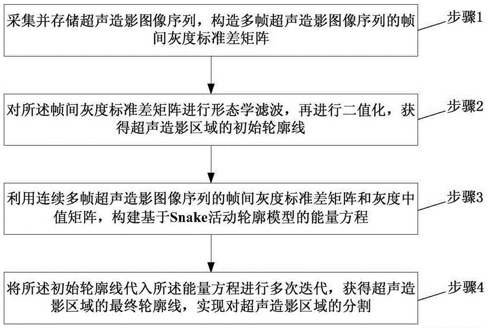
[0015] figure 1 It is a flowchart of a fully automatic ultrasound contrast image segmentation method according to an embodiment of the present invention. Such as figure 1 As shown, the method includes:

[0016] Step 1: Collect and store the contrast-enhanced ultrasound image sequence at a high frame rate, and construct an InterFrame Grayscale Standard Difference (IFGSD) matrix of the multi-frame contrast-enhanced ultrasound image sequence.

[0017] Step 2, performing a morphological filter on the inter-frame gray standard deviation matrix, and then performing binarization to obtain an initial contour line of the contrast-enhanced ultrasound region.

[0018] Specifically, step 1 and step 2 are to obtain the initial outline of the contrast region based on the int...


PUM


Abstract

The invention discloses a full-automatic contrast-enhanced ultrasonic image segmentation method. The method comprises the steps that step one, contrast-enhanced ultrasonic image sequences are acquired and stored, and an inter-frame grayscale standard difference matrix of multiple frames of contrast-enhanced ultrasonic image sequences is constructed; step 2, morphological filter is performed on the inter-frame grayscale standard difference matrix and then binarization is performed so that an initial contour line of an contrast-enhanced ultrasonic area is acquired; step three, an energy equation based on a Snake active contour model is constructed by utilizing the inter-frame grayscale standard difference matrix and the grayscale median matrix of continuous multiple frames of contrast-enhanced ultrasonic image sequences; and step 4, the initial contour line is substituted in the energy equation to perform multiple times of iteration so that the final contour line of the contrast-enhanced ultrasonic area is acquired, and segmentation of the contrast-enhanced ultrasonic area is realized.

Description

technical field [0001] The invention belongs to the technical field of ultrasound contrast imaging, in particular to a fully automatic ultrasound contrast image segmentation method. Background technique [0002] Ultrasound imaging is currently a relatively mature non-invasive detection method. Compared with CT and MRI (Magnetic Resonance Imaging, Magnetic Resonance Imaging, Magnetic Resonance Imaging, Magnetic Resonance Imaging), ultrasonic diagnosis has the advantages of less radiation, low cost, and convenient operation, making ultrasonic diagnosis the primary clinical examination method. Contrast-enhanced ultrasound technology uses contrast agents to enhance scattered echoes, which can effectively improve the resolution, sensitivity and specificity of ultrasound diagnosis. Therefore, contrast-enhanced ultrasound imaging has been hailed as the third revolution in ultrasound technology. At present, the imaging of contrast agent microbubbles by nonlinear detection method is...

Claims


Application Information

Patent Timeline
Patent Type & Authority Applications(China)
IPC IPC(8): G06T7/00
CPCG06T2207/10016G06T2207/10132G06T2207/30101
Inventor 钱明牛丽丽郑海荣周伟
Owner SHENZHEN INST OF ADVANCED TECH CHINESE ACAD OF SCI
Why Eureka
Try Eureka