
A 3D reconstruction method of intracranial nerve fiber bundles based on dti

A nerve fiber bundle and three-dimensional reconstruction technology is applied in the field of preparation of a three-dimensional solid model of the head, which can solve the problems of inability to provide three-dimensional intracranial anatomical structure and disease model images, and achieve the effect of reducing the risk of surgery.

Active Publication Date: 2019-09-20
MEDPRIN REGENERATIVE MEDICAL TECH
View PDF5 Cites 0 Cited by
  • Summary
  • Abstract
  • Description
  • Claims
  • Application Information

AI Technical Summary

Problems solved by technology

[0004] However, MRI and DTI can only provide local two-dimensional images for computer viewing, and cannot provide three-dimensional model images and / or models including intracranial anatomical structures and diseases

Method used


Image

  • A 3D reconstruction method of intracranial nerve fiber bundles based on dti
  • A 3D reconstruction method of intracranial nerve fiber bundles based on dti
  • A 3D reconstruction method of intracranial nerve fiber bundles based on dti

Examples


Embodiment Construction

[0058] The invention will be further described below in conjunction with the accompanying drawings, but the embodiments of the invention are not limited thereto.

[0059] In this embodiment, a test is conducted on a patient with a brain tumor, and a three-dimensional model including nerve fiber bundles, skull, brain tumor and blood vessels and a corresponding three-dimensional solid model are obtained through three-dimensional reconstruction and 3D printing.

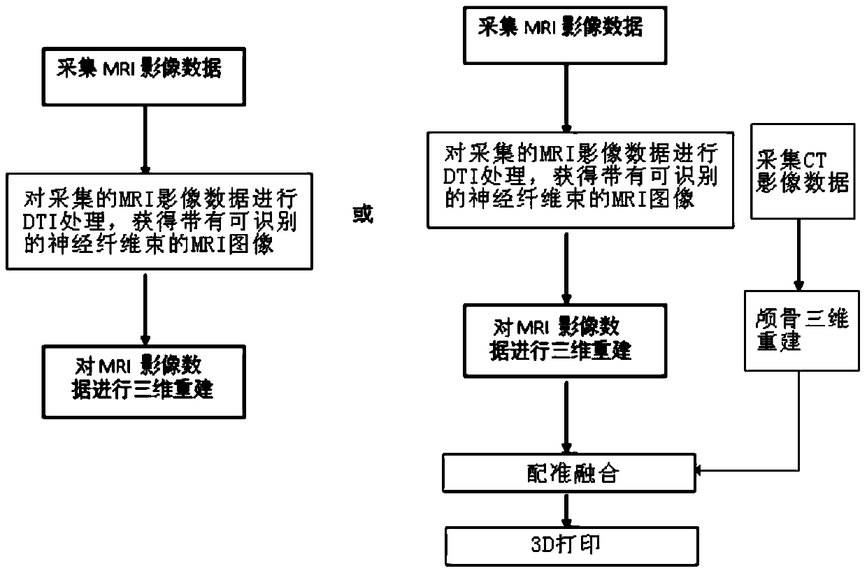
[0060] See attached figure 1 , a method for preparing a head three-dimensional solid model including nerve fiber bundles based on 3D printing technology, comprising the following steps:

[0061] S1: Use magnetic resonance scanning to scan the head lesion and related nerve fiber bundle area of ​​the brain tumor patient, and obtain the MRI image data of the head-related tissues, and the MRI image data is saved in DICOM format;

[0062] S2: Perform DTI processing on the acquired MRI images, including the calculation of dif...


PUM


Abstract

Provided are a DTI-based method for three-dimensional reconstruction of intracranial nerve fiber bundle, and a method for fabricating, on the basis of 3D printing technology, a skull three-dimensional model comprising nerve fiber bundles. Comprised are the following steps: performing magnetic resonance scanning of a target tissue area and a surrounding nerve fiber bundle to obtain MRI image data of the target tissue area containing the nerve fiber bundle; performing DTI- and related processing on the obtained MRI image and thereby obtaining an MRI image having a recognizable nerve fiber bundle; extrapolating the X-, Y-, and Z-axis information of the MRI image having the recognizable nerve fiber bundle, then three-dimensionally reconstructing by means of the Mimics software to obtain a three-dimensional model containing the intracranial nerve fiber bundle. By means of said three-dimensional model and the 3D-printed three-dimensional solid model, the positional relationships between anatomical structures, brain tissue functional areas, and the tissues can be clearly displayed, a solid model is provided for surgery and can be used for designing an operative route and simulating surgery.

Description

technical field [0001] The present invention relates to the technical field of three-dimensional reconstruction of images, more specifically, to a method for three-dimensional reconstruction of intracranial nerve fiber bundles based on DTI and a method for preparing a three-dimensional solid model of the head including nerve fiber bundles based on 3D printing technology . Background technique [0002] MRI (Magnetic Resonance Imaging) is an imaging technique that uses the signal generated by the resonance of atomic nuclei in a magnetic field to reconstruct the image. As a new medical imaging diagnostic technology, MRI has developed rapidly in recent years. The amount of information provided by magnetic resonance imaging is not only more than that of many other imaging techniques, but also has great potential advantages in diagnosing diseases with the unique information it provides. Patients with intracranial tumors need to undergo MRI examinations before surgery to diagnose...

Claims


Application Information

Patent Timeline
Patent Type & Authority Patents(China)
IPC IPC(8): G06T17/00
Inventor 兰青刘曼范小敏袁玉宇
Owner MEDPRIN REGENERATIVE MEDICAL TECH
Learn More