
Generalized side-lobe blanking method for ultrasonic system images

An ultrasound system and ultrasound image technology, which is applied in the field of medical equipment and signal processing, can solve the problems of no mature technical reports on the generalized sidelobe cancellation structure, and achieve the effect of improving the lateral resolution

Inactive Publication Date: 2016-07-06
TIANJIN UNIV
View PDF1 Cites 5 Cited by
  • Summary
  • Abstract
  • Description
  • Claims
  • Application Information

AI Technical Summary

Problems solved by technology

[0007] At present, there is no mature technical report on the application of generalized sidelobe cancellation structure in the field of ultrasound image processing

Method used


Image

  • Generalized side-lobe blanking method for ultrasonic system images
  • Generalized side-lobe blanking method for ultrasonic system images
  • Generalized side-lobe blanking method for ultrasonic system images

Examples


Embodiment Construction

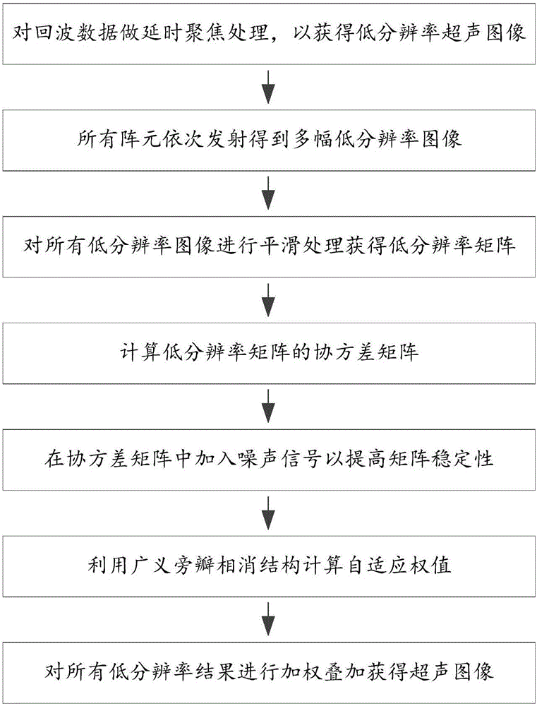
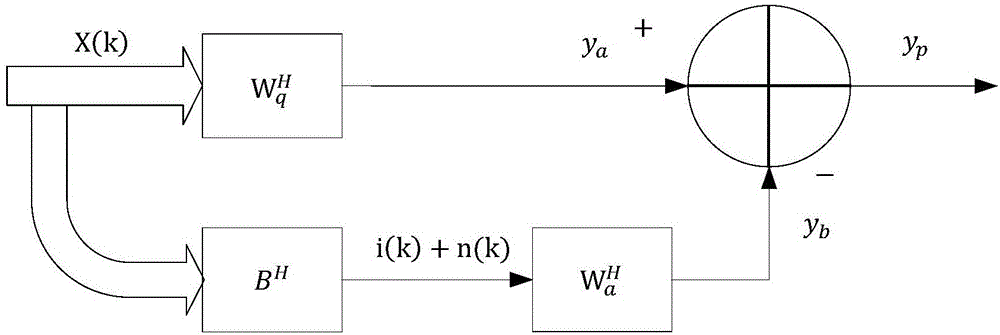
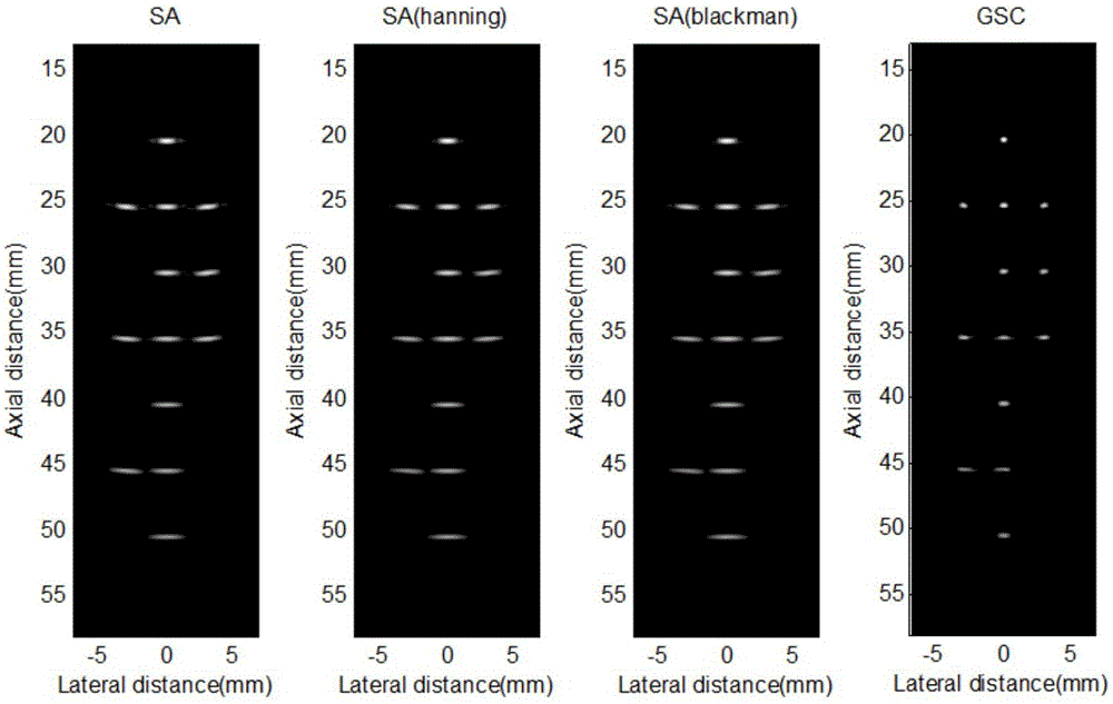
[0044] The purpose of the present invention is to provide a generalized sidelobe elimination method for medical ultrasound system to improve system resolution. The present invention uses time-delay focusing to obtain all low-resolution images, and smoothes all low-resolution images to obtain a low-resolution matrix and calculates its covariance matrix accordingly, combining the generalized sidelobe cancellation structure to combine the desired signal and noise The interference signal is distinguished, and the adaptive calculation weight matrix replaces the traditional non-adaptive weight, thus effectively improving the system resolution.

[0045] A generalized sidelobe elimination method for a medical ultrasound system, the method comprising:

[0046] Step 1. Use the single array element of the medical ultrasound transducer to emit ultrasonic waves, and use all array elements to receive ultrasonic echoes, and then perform time-delay focusing processing on the echo data to obta...


PUM


Abstract

The invention relates to medical instrument and signal processing technologies. For providing a generalized side-lobe blanking method for a medical ultrasonic system for improving the resolution of the system, the invention adopts the technical solution that a generalized side-lobe blanking method for ultrasonic system images comprises the steps of acquiring all low-resolution images using delay focusing, smoothing all the low-resolution images to obtain a low-resolution matrix, then calculating a covariance matrix, distinguishing desired signals from noise interference signals in combination with a generalized side-lobe blanking structure, and adaptively calculating a weight matrix to replace traditional non-adaptive weights, so as to improve the resolution of ultrasonic images. The method is mainly applied to image processing.

Description

technical field [0001] The invention relates to medical equipment and signal processing technology, in particular to data processing technology in medical ultrasonic imaging, especially a method for improving the resolution of an ultrasonic system. Background technique [0002] Ultrasound imaging has the characteristics of strong transmission ability and little harm to the human body, and has become one of the most important medical diagnostic techniques. Ultrasonic imaging is a method and technology that uses ultrasonic waves to irradiate a test object, and obtains visible images of the tissue properties and structures of the test object by receiving and processing echoes that carry characteristic information of the internal tissue or structure properties of the test object. Ultrasound imaging has the advantages of low price, good real-time imaging, and no harm to patients. [0003] The resolution of ultrasonic images is one of the main performances of ultrasonic diagnosti...

Claims


Application Information

Patent Timeline
IPC IPC(8): G06T3/40G06T5/00G06T7/00
CPCG06T3/4076G06T7/0012G06T2207/10132G06T5/70
Inventor 陈晓冬时一峰李嘉科汪毅郁道银
Owner TIANJIN UNIV
Learn More