Tumor region image enhancement method and system based on enhanced composite image

A tumor area and enhanced image technology, applied in the field of medical image processing, can solve problems such as low accuracy, fuzzy tumor boundaries, and difficult tumor boundary segmentation

Inactive Publication Date: 2016-07-06
SOUTH CENTRAL UNIVERSITY FOR NATIONALITIES
View PDF6 Cites 17 Cited by
  • Summary
  • Abstract
  • Description
  • Claims
  • Application Information

AI Technical Summary

Problems solved by technology

Accurate segmentation of tumor boundaries is very important for treatment planning. At present, this part of work mainly relies on manual delineation, and the accuracy is not high
Due to the noise and ambiguity in medical images such as CT or MR, t

Method used


Image

  • Tumor region image enhancement method and system based on enhanced composite image
  • Tumor region image enhancement method and system based on enhanced composite image
  • Tumor region image enhancement method and system based on enhanced composite image

Examples


Embodiment Construction

[0079] The present invention will be further described in detail below in conjunction with the accompanying drawings and specific embodiments.

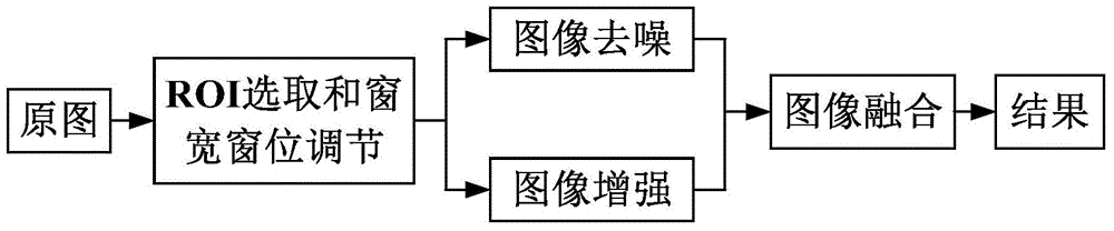
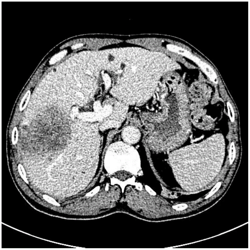
[0080] Aiming at the problem that the boundary of the tumor in the original CT or MR image is blurred and difficult to segment, an embodiment of the present invention provides an image enhancement method for a tumor region based on a synthetic enhanced image, see figure 1 As shown, the method includes the following steps:

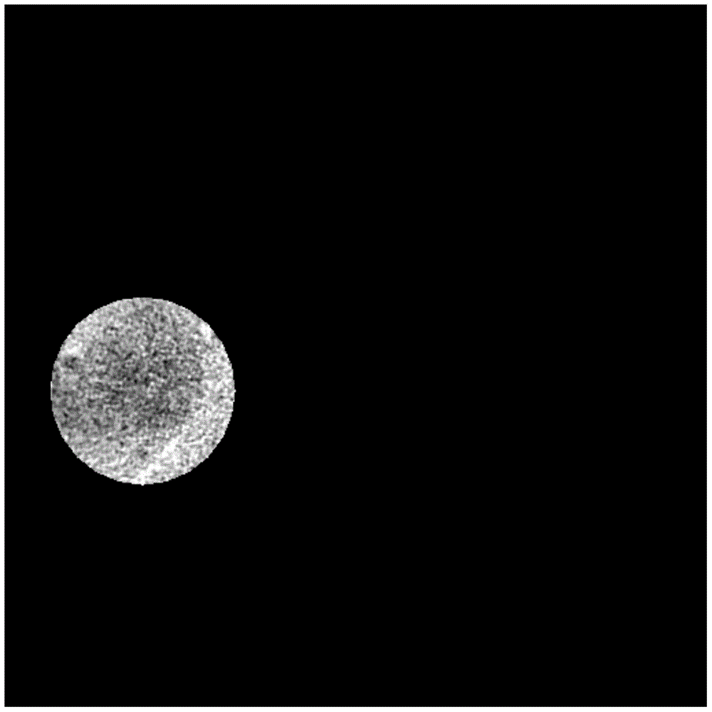
[0081] A. In an original CT image or MR image containing a tumor, select an elliptical ROI (RegionOfInterest, region of interest) covering the entire tumor area; set all pixels outside the ROI area to zero, and set all pixels outside the ROI area to zero. The pixel value is used to adjust the window width and window level to meet the requirements of human observation.

[0082] In practical applications, the ROI area is first selected, and then the window width and level are adjusted. However, since the pixel value...


PUM


Abstract

The invention discloses a tumor region image enhancement method and system based on an enhanced composite image, and relates to the field of medical image processing. The invention puts forwards a concept "enhanced composite image" for the first time, a ROI (Region Of Interest) which covers all tumor regions is independently subjected to noise reduction processing and enhancement processing in an original CT (Computed Tomography) or MR (Magnetic Resonance) image to obtain a noise reduction image and an enhancement image of the RIO, and the noise reduction image and the enhancement image are subjected to weight fusion to obtain the enhanced composite image. The RIO in the original CT or MR image can be enhanced, and different weighting factors in the enhanced composite image are selected to cause the surface and the fuzzy boundary of the tumor in the CT or MR image to become clear so as to bring convenience for doctors to observe the surface and the boundary of the tumor. A threshold value segmentation method is adopted on the enhanced composite image, and therefore, the tumor region can be precisely segmented.

Description

technical field [0001] The present invention relates to the field of medical image processing, in particular to a tumor region image enhancement method and system based on synthetically enhanced images. Background technique [0002] In the process of actually diagnosing tumor-related diseases, doctors can help clinical patients by measuring and analyzing the boundary, cross-sectional area and volume of tumors in medical images such as CT (Computed Tomography) or MR (Magnetic Resonance, magnetic resonance). To diagnose the disease, it is necessary to segment the tumor border in medical images such as CT or MR. Accurate segmentation of tumor boundaries is very important for treatment planning. At present, this part of work mainly relies on manual delineation, and the accuracy is not high. Due to the noise and ambiguity in medical images such as CT or MR, the boundaries of many tumors are blurred. Traditional image segmentation methods, such as threshold segmentation methods o...

Claims


Application Information

Patent Timeline
IPC IPC(8): G06T5/00G06T7/00
CPCG06T5/001G06T5/002G06T2207/20104G06T2207/30096
Inventor 谌先敢刘海华高智勇陆雪松
Owner SOUTH CENTRAL UNIVERSITY FOR NATIONALITIES
Why Eureka
Try Eureka