High resolution 3D phase microscopy imaging device and imaging method

A high-resolution, phase microscopy technique, applied in measurement devices, optical devices, instruments, etc., can solve the problems of impossible samples, inability to detect the refractive index of the sample, that is, the phase distribution, and slow imaging speed.

Active Publication Date: 2016-07-27
SHANGHAI INST OF OPTICS & FINE MECHANICS CHINESE ACAD OF SCI
View PDF5 Cites 11 Cited by
  • Summary
  • Abstract
  • Description
  • Claims
  • Application Information

AI Technical Summary

Problems solved by technology

Both coherent tomography and laser confocal illumination microscopy are based on three-dimensional point-by-point illumination to obtain three-dimensional images, so the imaging speed is relatively slow
Both have an irreparable defect, that is, in most cases, the phase information inside the sample is simply assumed to be uniformly distributed, which is impossible for actual samples
Therefore, all three-dimensional imaging techniques so far cannot detect the refractive index or phase distribution inside the sample.

Method used


Image

  • High resolution 3D phase microscopy imaging device and imaging method
  • High resolution 3D phase microscopy imaging device and imaging method
  • High resolution 3D phase microscopy imaging device and imaging method

Examples


Embodiment Construction

[0066] The present invention will be further described below in conjunction with the embodiments and accompanying drawings, but the protection scope of the present invention should not be limited thereby.

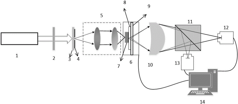
[0067] see first figure 1 , figure 1 It is a diagram of the high-resolution three-dimensional phase microscopic imaging device of the present invention. It can be seen from the figure that the high-resolution three-dimensional phase microscopic imaging device of the present invention includes a coherent light source 1, a two-dimensional electric translation stage 6 moving perpendicular to the optical axis, a first detector 12 and a computer 14, and is characterized in that there are also attenuation plates 2, Ground glass 3, pinhole diaphragm 4, 4F zoom system 5, standard glass plate 8, resolution plate 9, micromagnification system 10, beam splitting prism 11 and second detector 13, the positional relationship of the above-mentioned components is as follows:

[0068] Al...


PUM


Abstract

The invention relates to a high resolution 3D phase microscopy imaging device and an imaging method, a scattering light spot illumination sample limited by a small aperture is adopted, a micro-amplifier system is adopted to amplify a diffraction spot behind a to-be-detected element. A virtual detector concept is adopted on data processing, the to-be-detected element is seen as a 3D body formed by multiple sections, the iterative operation is performed between the multiple sections and the virtual detector, updated layers are added, complex amplitude transmittance function and illumination light distribution of corresponding layers are updated, the phase distribution of section of each layer of the to-be-detected element is obtained at last, a cross section image interpolation is combined with the above distribution, 3D phase imaging of the to-be-detected element is obtained. The technology is only one 3D microscopy imaging technology which can detect the refractive index of internal element, that is, phase distribution condition at present.

Description

technical field [0001] The invention relates to three-dimensional microscopic imaging, in particular to a high-resolution three-dimensional phase microscopic imaging device and imaging method. Background technique [0002] The three-dimensional microscopic imaging technologies in the traditional optical field mainly include coherent tomography and confocal scanning microscopic imaging. Both coherent tomography and laser confocal illumination microscopy are based on three-dimensional point-by-point illumination to obtain three-dimensional images, so the imaging speed is relatively slow. Both have an irreparable defect, that is, in most cases, the phase information inside the sample is simply assumed to be uniformly distributed, which is impossible for actual samples. Therefore, all three-dimensional imaging techniques so far cannot detect the refractive index, that is, the phase distribution inside the sample. [0003] In 2015, based on the ePIE algorithm (see MaidenAM, Rod...

Claims


Application Information

Patent Timeline
Patent Type & Authority Applications(China)
IPC IPC(8): G01B11/24
CPCG01B11/24
Inventor 刘诚陈文王海燕朱健强
Owner SHANGHAI INST OF OPTICS & FINE MECHANICS CHINESE ACAD OF SCI
Why Eureka
Try Eureka