Specific detection and quantification of cardiolipin and isolated mitochondria by positively charged AIE fluorogens and method of manufacturing of AIE fluorogens

A mitochondrial and cardiolipin technology, applied in fluorescence/phosphorescence, chemical instruments and methods, measurement devices, etc., can solve problems such as poor water solubility, unclear working mechanism, and difficulty in use

Active Publication Date: 2016-08-17
THE HONG KONG UNIV OF SCI & TECH
View PDF13 Cites 14 Cited by
  • Summary
  • Abstract
  • Description
  • Claims
  • Application Information

AI Technical Summary

Problems solved by technology

In addition, the small Stokes shift and poor water solubility of NAO make it difficult to use in biological systems
How NAO works is unclear and performance is difficult to improve, even wit

Method used


Image

  • Specific detection and quantification of cardiolipin and isolated mitochondria by positively charged AIE fluorogens and method of manufacturing of AIE fluorogens
  • Specific detection and quantification of cardiolipin and isolated mitochondria by positively charged AIE fluorogens and method of manufacturing of AIE fluorogens
  • Specific detection and quantification of cardiolipin and isolated mitochondria by positively charged AIE fluorogens and method of manufacturing of AIE fluorogens

Examples


Embodiment Construction

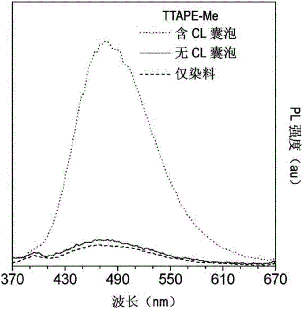
[0039] The inventors discovered that certain AIE luminophores can be used to quantify cardiolipin and isolate mitochondria. More specifically, these AIE luminophores can be applied to samples, cells, or vesicles, allowing the cells to be imaged. This allows quantification of cardiolipin or isolation of mitochondria.

[0040] The AIE luminophore may be the fluorophore TTAPE-ME. The TTAPE-ME can bind lipobinding protein, which can be a lipobinding fragment of any protein.

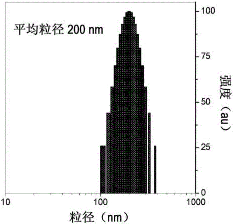
[0041] Cardiolipin can be located in the cytosol or membrane. The membrane may be in the form of a cell membrane or a lipid vesicle. The lipid vesicle may be a large unilamellar vesicle (LUV), i.e. the vesicle has a diameter of about 60nm to 800nm, 70 to 800nm, 80nm to 800nm, 90nm to 800nm, 100nm to 700nm, 200nm to 600nm, 300nm to 500nm, 400nm to 800nm, 500nm to 800nm, 600nm to 800nm, or 700nm to 800nm.

[0042] The cell membrane may be a eukaryotic cell membrane. The eukaryotic cell membrane may be a m...


PUM


Abstract

The present subject matter relates to a one-step method of detecting and quantifying cardiolipin in a sample using a positively charged AIE luminogen by introducing the AIE luminogen to a solution containing the sample and measuring fluorescence intensity of the solution; a method of quantifying isolated mitochondria using a positively charged AIE luminogen by staining a sample containing isolated mitochondria with the AIE luminogen and measuring the fluorescence intensity; and a method of quantifying isolated mitochondria using a positively charged AIE luminogen by introducing the AIE luminogen to a sample containing isolated mitochondria, wherein the AIE luminogen stains the isolated mitochondria and identifying the stained isolated mitochondria under microscope. With improved sensitivity and excellent selectivity to CL over other major mitochondrial membrane lipids, an aggregation-induced emission-active fluorogen, TTAPE-Me, may serve as a valuable fluorescent sensor for CL detection and quantification and the quantification of isolated mitochondria.

Description

[0001] Cross References to Related Applications [0002] This patent application claims priority to U.S. Provisional Patent Application No. 61 / 963,393, filed December 3, 2013, the applicant of which is the inventor of this patent application, the entire contents of which are incorporated herein by reference . technical field [0003] The subject of the present invention relates in particular to compounds or salts which allow the quantification of cardiolipin (CL) and which allow the quantification and identification of isolated mitochondria, in particular from the yeast S. cerevisiae strain YPH 500. Background technique [0004] Eukaryotic cells use about 5% of their genes to synthesize lipids. Due to the indispensable functions of lipids in cells, a large fraction of lipids is utilized. Due to their unique structure, lipids form bilayer structures to separate internal components from the extracellular environment and to compartmentalize individual cellular organelles. In...

Claims


Application Information

Patent Timeline
IPC IPC(8): G01N21/64C09K11/06
CPCG01N21/6458G01N21/6428G01N33/483
Inventor 唐本忠洪煜柠梁玮彤
Owner THE HONG KONG UNIV OF SCI & TECH
Why Eureka
Try Eureka