Group of colon cancer metastasis related tumor stroma markers and application

A technology of tumor stroma and markers, applied in the biological field, can solve the problems of insufficient specificity of diagnostic molecules and inaccurate early diagnosis

Inactive Publication Date: 2016-08-24
XIANGYA HOSPITAL CENT SOUTH UNIV
View PDF4 Cites 3 Cited by
  • Summary
  • Abstract
  • Description
  • Claims
  • Application Information

AI Technical Summary

Problems solved by technology

[0004] Therefore, the present invention aims to overcome the problem that the diagnostic molecules in the prior art are not specific enough and can not be accurately and early diagnosed

Method used


Image

  • Group of colon cancer metastasis related tumor stroma markers and application
  • Group of colon cancer metastasis related tumor stroma markers and application
  • Group of colon cancer metastasis related tumor stroma markers and application

Examples


Embodiment 1

[0020] Analysis of Differentially Expressed Proteins in Carcinogenesis of Colon Cancer

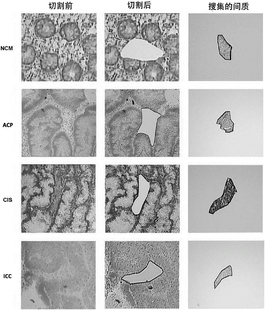
[0021] Collect the pathological tissues of the four typical stages (normal, adenomatous polyps, carcinoma in situ, and invasive carcinoma) in the process of colon cancer carcinogenesis. After the section was stained, it was confirmed by a pathologist. After the tissue was frozen sectioned, it was pasted on a special membrane for microdissection, fixed in 75% ethanol solution for 30 seconds, and stained with 0.5% methyl green. After staining, the sections are air-dried for later use. After staining, the sections were subjected to laser microdissection. Under the microscope, select areas with a consistency greater than 95% for cutting, and collect cutting components.

[0022] The result is figure 1 As shown, by microdissection, the interstitial tissue surrounding colon epithelial cells is effectively separated and collected. This shows that the subsequent identified proteins all come from int...

Embodiment 2

[0024] Extract the collected interstitial components at different stages: dissolve the interstitial components in an appropriate amount of lysis buffer (7MUrea, 2M Thiourea, 65mM DTT, 0.1mM PMSF) and lyse at 4℃ for 1 hour, then centrifuge at 12000rpm for 30 minutes Collect the supernatant. A 2D quantitative kit was used for protein concentration determination. Different individual samples at the same stage are mixed in equal amounts to form four-stage mixed protein samples for subsequent labeling experiments.

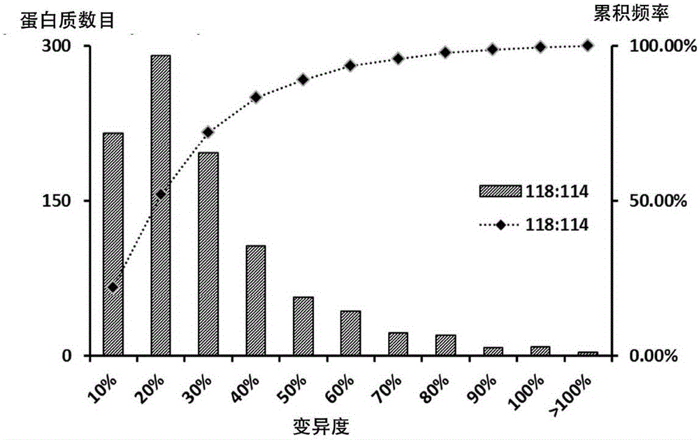
[0025] The samples were labeled according to the standard procedure of the iTRAQ kit: 4 mixed protein samples, each of which was 100 ug, was reduced and alkylated and then digested overnight at 37°C. The peptides after enzymolysis were labeled with 8-channel (8plex) iTRAQ reagent (labeling reagent 114,118 labeled NCM interstitial; reagent 113,117 labeled ACP interstitial; reagent 115,119 labeled CIS interstitial; reagent 116,121 labeled ICC interstitial). After labeling,...

Embodiment 3

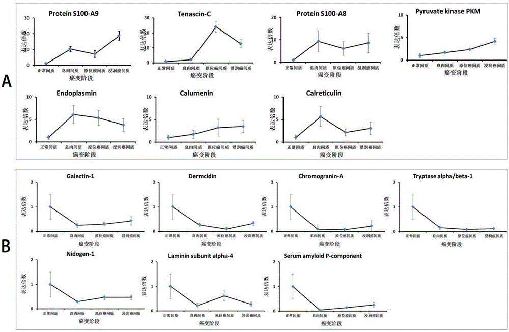
[0030] Tenascin-C expression in different stages of tissues (see Table 2)

[0031] Collected 50 cases of NCM, 50 cases of ACP, 30 cases of CIS and 63 cases of ICC paraffin-embedded tissue sections. After the sections were deparaffinized, they were incubated with Tenascin-C antibody (1:1000) overnight, and after reaction with peroxidase-labeled secondary antibodies, DAB developed color. The sections were counterstained with hematoxylin. Score the positive density and staining intensity of each slice under the microscope. The scoring range is 0-6 seven levels: 0, 1, 2 are no expression; 3, 4 are low expression, 5, 6 are high expression. The staining result is like Figure 4 Shown.

[0032] The results are shown in Table 2: 74% of the normal mesenchyme has no expression, 22% has low expression, and only 2% has high expression. However, only 16% of infiltrating tumor stroma has low expression and 45% high expression. Statistics show that the difference is significant. It shows tha...


PUM


Abstract

The invention discloses a group of colon cancer metastasis related tumor stroma markers. The group of marks exist in a tumor stroma, and marker expression abnormality is also more obvious along with the development of canceration, wherein one marker Tenascin-C belongs to a novel extracellular matrix marker, generates abnormal expression in the early stage of tumor canceration, and also generates abnormal expression in a metastasis position, wherein the marker for the self secretion of a tumor is relevant to the metastasis capability. The group of markers can be used for preparing medicines and a kit for colon cancer metastasis screening and auxiliary diagnosis.

Description

Technical field [0001] The invention belongs to the field of biotechnology, and specifically relates to a group of colon cancer metastasis-related tumor interstitial markers and applications. Background technique [0002] Colon cancer (CC) is one of the most common malignant tumors in the gastrointestinal tract, and its incidence has increased significantly in recent years. More than one million people are deeply affected by it every year around the world, and most of them are in the middle or late stage when they are discovered, which seriously threatens human health. The occurrence and metastasis of tumors are closely related to the microenvironment of tumor cells. Therefore, screening key proteins from the microenvironment in the process of colon cancer carcinogenesis is of great significance for in-depth understanding of the mechanism of carcinogenesis, finding diagnostic and therapeutic targets, and preventing tumorigenesis. [0003] At present, the diagnosis and treatment o...

Claims


Application Information

Patent Timeline
Patent Type & Authority Applications(China)
IPC IPC(8): G01N33/574
CPCG01N33/57484G01N33/57419
Inventor 李茂玉陈永恒陈主初彭芳黄英
Owner XIANGYA HOSPITAL CENT SOUTH UNIV
Why Eureka
Try Eureka