
Single-excitation fat-water separation imaging error correction system and method
A technology for correcting system and imaging errors, applied in measuring devices, instruments, measuring electrical variables, etc., can solve problems such as background noise interference, phase winding artifacts, and the time required for several seconds to tens of seconds
- Summary
- Abstract
- Description
- Claims
- Application Information
AI Technical Summary
Problems solved by technology
Method used
Image

Examples
Embodiment 1
[0163] On the 0.35T medical magnetic resonance imager, set the center frequency of the radio frequency pulse to the water proton resonance frequency (14.9MHz), press Figure eight The workflow shown executes the modules.
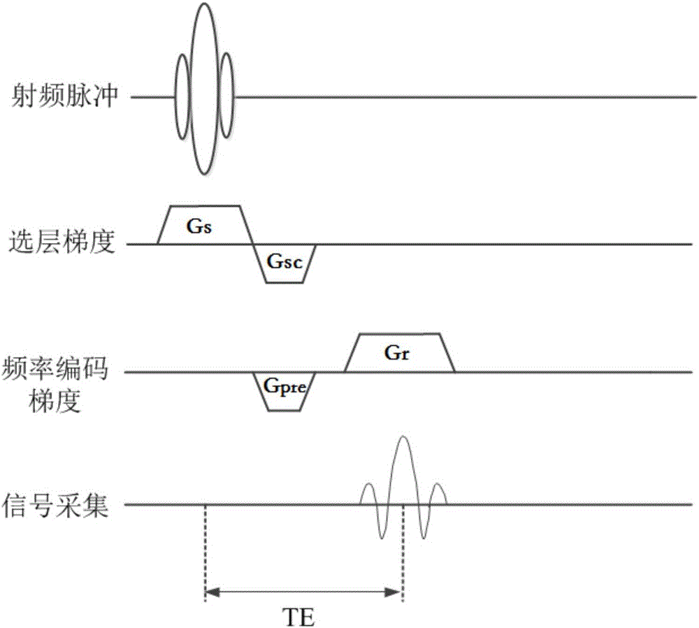
[0164] First, run the pre-scan module, locate the signal acquisition area within the area of interest of the human body, and run Figure 1 In the apparent transverse relaxation time test sequence shown, the data preprocessing module obtains the apparent transverse relaxation time constant through nonlinear fitting of the echo amplitudes of different TEs based on formula (1) When the eddy current effect is significant, the pre-scan module performs gradient waveform pre-emphasis compensation in the following manner:
[0165] A. The positioning signal acquisition area is located within the area of interest of the human body. For Figure six The eddy current test sequence shown, sets the amplitude, width, and polarity of the read gradient pulse with the ...
Embodiment 2
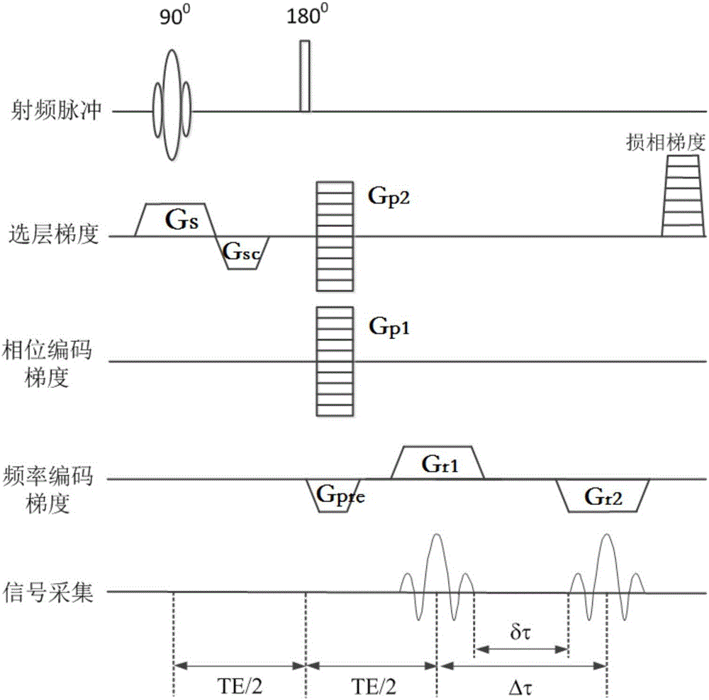
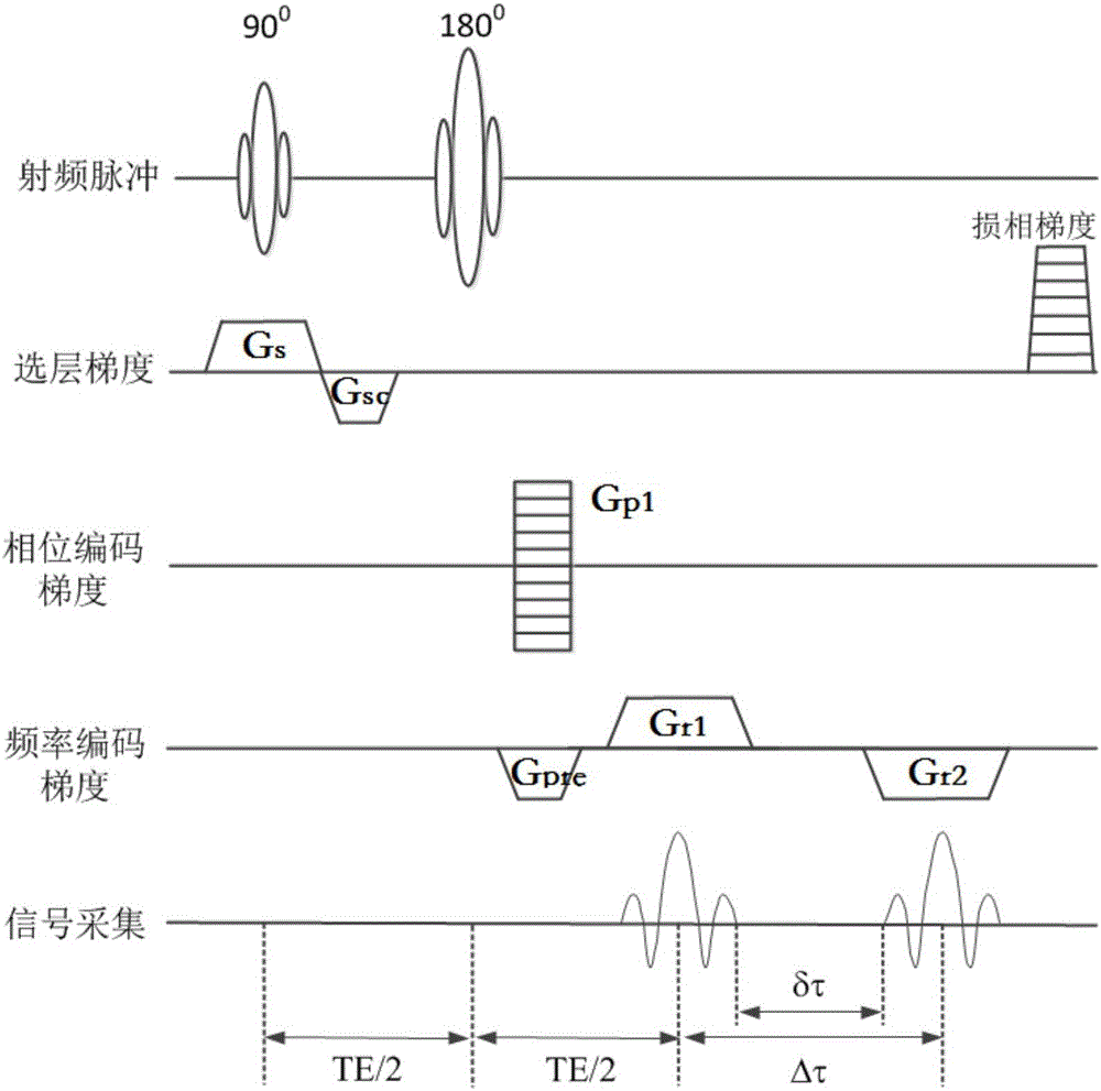
[0186] On the 1.5T medical magnetic resonance imager, set the center frequency of the radio frequency pulse as the proton resonance frequency (63.8MHz), according to Figure 8 The workflow shown executes each module, starting with the pre-scan module run Figure 1 For the apparent transverse relaxation time test sequence shown, the data preprocessing module obtains the apparent transverse relaxation time constant + Then, the hydrolipid scan module runs Figure II In the three-dimensional version of the dual gradient echo chemical shift imaging sequence shown, the integral area of the first frequency encoding gradient is set to be twice the integral area of the pre-reading gradient in the sequence. The two polarities are opposite, and the second frequency encoding is set The polarity of the gradient is opposite to that of the first frequency encoding gradient, and the integral areas of the two are equal. The 180° hard pulse width is calibrated to 100 μs, and the time inte...
PUM
 Abstract
Description
Claims
Application Information