Method, device and cell analyzer for counting white blood cells

A white blood cell count and white blood cell technology, applied in the field of cell analyzers, can solve the problems of false increase of white blood cell count value, affecting clinical judgment, etc.

Active Publication Date: 2020-11-13
SHENZHEN MINDRAY BIO MEDICAL ELECTRONICS CO LTD
View PDF8 Cites 0 Cited by
  • Summary
  • Abstract
  • Description
  • Claims
  • Application Information

AI Technical Summary

Problems solved by technology

However, when there are lipid particles in the blood sample, such as after injecting fat emulsion, using the BASO channel to count the white blood cells, there will be a false increase in the white blood cell count, which will affect the clinical judgment

Method used


Image

  • Method, device and cell analyzer for counting white blood cells
  • Method, device and cell analyzer for counting white blood cells
  • Method, device and cell analyzer for counting white blood cells

Examples


Embodiment 1

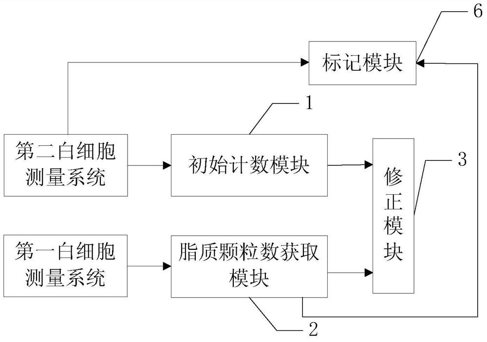
[0045] Based on the above white blood cell counting device, this implementation also discloses a white blood cell counting method, please refer to figure 2 , is a flow chart of the leukocyte counting method, and the method includes the following steps:

[0046] Step S100, initial counting. The initial white blood cell count value is counted from the second white blood cell measurement system of the cell analyzer, wherein the second white blood cell measurement system is a measurement system that can at least classify the white blood cells into basophils and other white blood cells, and collects the first count of cells flowing through the detection area. Scattered light signals and side scattered light signals form a scatter diagram. In a specific embodiment, the initial count value can be the number of particles in the non-ghost area in the scatter diagram of the second white blood cell measurement system; in another specific embodiment, the initial count value can also be ...

Embodiment 2

[0074] Please refer to figure 2 , image 3 and Figure 7 , the white blood cell counting method disclosed in this embodiment also includes marking the lipid particles in the scatter diagram of the second white blood cell measurement system, including:

[0075] Step S400, lipid particle labeling. Please refer to Figure 8, usually, the cell analyzer will present the scattergrams of the first and second white blood cell measurement systems on the display interface of the display device in a visualized form. In this embodiment, the visually displayed scatter diagram at least includes a two-dimensional scatter diagram of side scattered light-forward scattered light of the second white blood cell measurement system. In other embodiments, it can also display Other scatterplots, such as adding dimensions or displaying other scatterplots. In order to improve the user experience, the user can visually see the distribution of lipid particles in the white blood cell area on the sca...

Embodiment 3

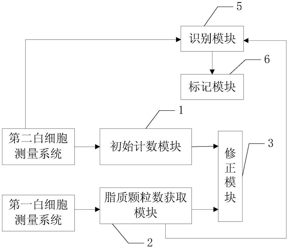
[0079] The white blood cell counting method disclosed in this embodiment also includes: identifying lipid particles in the scattergram of the second white blood cell measurement system, please refer to Figure 9 , which is a flow chart of identifying lipid particles in this embodiment, specifically includes the following steps:

[0080] Step S510, dividing the first sub-region. In the scatter diagram of the first white blood cell measurement system, the scatter diagram is divided into several first sub-regions along the direction of the side scattered light. Please refer to Figure 10a , 10b and 10c, in a specific embodiment, can be equally divided into N segments along the direction of side scattered light, N is 5 to 20, preferably N=8, the specific value can be determined according to the resolution of the scatter diagram, the resolution High, N can be larger. In other embodiments, it may also be divided into N segments unequally.

[0081] Step S520, dividing the second...


PUM


Abstract

The invention provides a white blood cell count method and a white blood cell count device. The white blood cell initial count value is counted by a second white blood cell measuring system of a cell analyzer; and the initial count value is corrected by a lipid granule number obtained from a first white blood cell measuring system, so that the interference of the lipid granules in the second white blood cell measuring system on the white blood cell count is reduced, and the white blood cells are accurately counted. According to the white blood cell count method and the white blood cell count device, the cell analyzer is also provided.

Description

technical field [0001] The invention relates to the field of cell analyzers, in particular to a white blood cell counting method, device and cell analyzer. Background technique [0002] Hematology analyzer is an instrument that can detect cells in blood. It can count and classify white blood cells (WBC), red blood cells, platelets, nucleated red blood cells, reticulocytes and other cells. [0003] One of the most common methods for blood cell analyzers to detect white blood cells is the laser scattering method, which uses laser light to irradiate the cells flowing through the detection area, and collects the light signals reflected or scattered by the cells to classify and count white blood cells. Optical signals include forward scattered light, side scattered light, fluorescence, etc. The forward scattered light signal reflects the size information of the cell, the side scattered light signal reflects the complexity of the internal structure of the cell, and the fluorescen...

Claims


Application Information

Patent Timeline
Patent Type & Authority Patents(China)
IPC IPC(8): C12Q1/06C12M1/34
Inventor 叶波王官振郑文波
Owner SHENZHEN MINDRAY BIO MEDICAL ELECTRONICS CO LTD
Why Eureka
Try Eureka