Ear-nose-throat endoscope equipment

An endoscope and ear, nose and throat technology, applied in the field of ear, nose and throat endoscope equipment, can solve the problems of poor bendability, too large outer diameter of the mirror tube, and unclear images, so as to enhance the flexible bending performance and ensure the treatment Time, improve the effect of long-acting

Inactive Publication Date: 2016-10-26
张学军
View PDF9 Cites 8 Cited by
  • Summary
  • Abstract
  • Description
  • Claims
  • Application Information

AI Technical Summary

Problems solved by technology

[0003] But it has the following disadvantages: 1. Due to the large outer diameter of the mirror tube and poor bendability, it cannot freely and flexibly enter and exit the ENT cavity
[0004] 2. The usual ENT endoscopic equipment is that the doctor holds the handle with one hand, and the other hand assists in dilating the patient's ENT, which cannot provide necessary treatment and is extremely inconvenient to use
[0005] 3. There is a large amount of human body water vapor in the ear, nose and throat cavity. The surface of traditional endoscopes is easy to condense water vapor and form a fog layer. Therefore, the usual practice is to soak the mirror body with physiological saline heated to about 45°C for 20 minutes before use. After entering the human body for observation, but after a long time, fogging cannot be avoided after the temperature of the lens body drops
[0006] 4. The common ENT endoscope equipment cannot zoom in on the lesion, which brings a lot of inconvenience to the medical staff.
[0007] 5. In order to see a clear image in the body, it is usually necessary to illuminate the environment in the body. At present, point light sources are generally used for illumination, and the light efficiency is low, resulting in the observed image not being clear enough, which affects the doctor's diagnosis of the patient's condition
[0008] 6. Due to the differences in the level of medical treatment in various regions, doctors in some underdeveloped areas may not be able to accurately determine the exact disease of the patient's ear, nose and throat, thus delaying the best treatment time for the patient

Method used


Image

  • Ear-nose-throat endoscope equipment
  • Ear-nose-throat endoscope equipment
  • Ear-nose-throat endoscope equipment

Examples


Embodiment Construction

[0024] The present invention will be described in further detail below in conjunction with the accompanying drawings and specific embodiments.

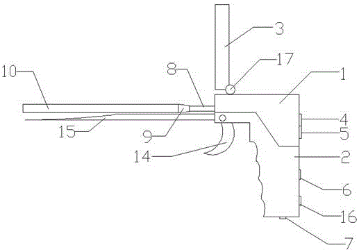
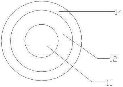
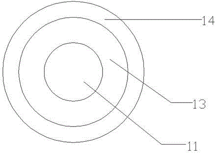
[0025] Such as Figure 1 ~ Figure 4 As shown in the structural schematic diagram of the present invention, a kind of otolaryngology endoscopic equipment, it comprises camera host 1 and handle 2, and described camera host 1 is provided with described camera host 1 at one end of described handle 2; Described camera host 1 is provided with A liquid crystal display 3; the camera host 1 is provided with a wireless transmitting and receiving module; one side of the camera host 1 is provided with a shooting switch 4 and a camera switch 5; the camera host 1 is provided with a camera host socket 8; The camera host socket end 8 is provided with a mirror tube interface 9; one side of the mirror tube interface 9 is provided with a mirror tube 10; the mirror tube interface 9 and the mirror tube 10 are integrally formed; the mirror The inner wall ...


PUM


Abstract

The invention discloses ear-nose-throat endoscope equipment. The ear-nose-throat endoscope equipment comprises a camera host (1) and a hand-held handle (2), wherein the camera host is arranged at one end of the hand-held handle; the camera host is provided with a liquid crystal display screen (3); a wireless transmitting-receiving module is arranged in the camera host; a shooting switch (4) and a photographing switch (5) are arranged on one side of the camera host; a camera host socket end (8) is arranged on the camera host; an endoscope pipe interface end (9) is arranged on the camera host socket end; an endoscope pipe (10) is arranged on one side of the endoscope pipe interface end; an endoscope (11) and an annular heating ring (12) are arranged on the inner wall at one end of the endoscope pipe; the hand-held handle is provided with an expansion trigger (14); and the expansion trigger is connected with a disposable expander or a tongue depressor (15) though the hand-held handle. The endoscope pipe adopts medical silica gel or an anti-microbial nanomaterial; the flexibility and bending performance of the endoscope pipe are reinforced due to the medical silica gel; the anti-microbial nanomaterial has the long-term anti-microbial and sterilizing function due to the slow release effect of the anti-microbial nanomaterial, so that the anti-microbial and sterilizing long-term effect of the ear-nose-throat endoscope equipment is improved.

Description

technical field [0001] The invention relates to the technical field of medical instruments, in particular to an ENT endoscope device. Background technique [0002] In the medical field, medical diagnosis using an endoscope has been widely used. Specifically, an endoscope and a light source are built into one end of the mirror tube inserted into the body cavity to take pictures of the lesion in the body cavity, and then display the image on the display screen for the doctor to observe for diagnosis. For example, in the process of diagnosing ENT-related diseases, it is necessary to insert an endoscope-equipped mirror tube into the patient's ENT for inspection, and to take images of the inside of the patient's ENT through the endoscope. The image is sent to the LCD screen. [0003] But it has the following disadvantages: 1. Due to the excessively large external diameter of the mirror tube and poor bendability, it cannot freely and flexibly enter and exit the ENT cavity. [0...

Claims


Application Information

Patent Timeline
Patent Type & Authority Applications(China)
IPC IPC(8): A61B1/227A61B1/233A61B1/267A61B1/253A61B1/06A61B1/04A61B1/00
CPCA61B1/227A61B1/00009A61B1/00016A61B1/00064A61B1/00103A61B1/00131A61B1/04A61B1/0607A61B1/0684A61B1/233A61B1/253A61B1/267
Inventor 张学军
Owner 张学军
Why Eureka
Try Eureka