
A super-resolution optical imaging method based on a single-molecule positioning process

An optical imaging and super-resolution technology, applied in the field of biophotonics technology, can solve the problems of inability to observe exosomes in detail, and achieve the effects of targeted observation and imaging, high labeling density, and high yield of fluorescent molecules

Active Publication Date: 2016-10-26
SOUTHEAST UNIV
View PDF5 Cites 10 Cited by
  • Summary
  • Abstract
  • Description
  • Claims
  • Application Information

AI Technical Summary

Problems solved by technology

[0004] Purpose of the invention: In order to overcome the deficiencies in the prior art, the present invention provides a super-resolution optical imaging method based on single-molecule localization to observe the interaction process between tumor cell exosomes and normal cells. In the existing technology, due to the limitation of diffraction limit, it is impossible to observe exosomes in detail, which provides a new technical means for studying the mechanism of cancer metastasis mediated by exosomes and the treatment of cancer metastasis and diffusion.

Method used


Image

  • A super-resolution optical imaging method based on a single-molecule positioning process

Examples


Embodiment Construction

[0022] Below in conjunction with embodiment the present invention will be further described.

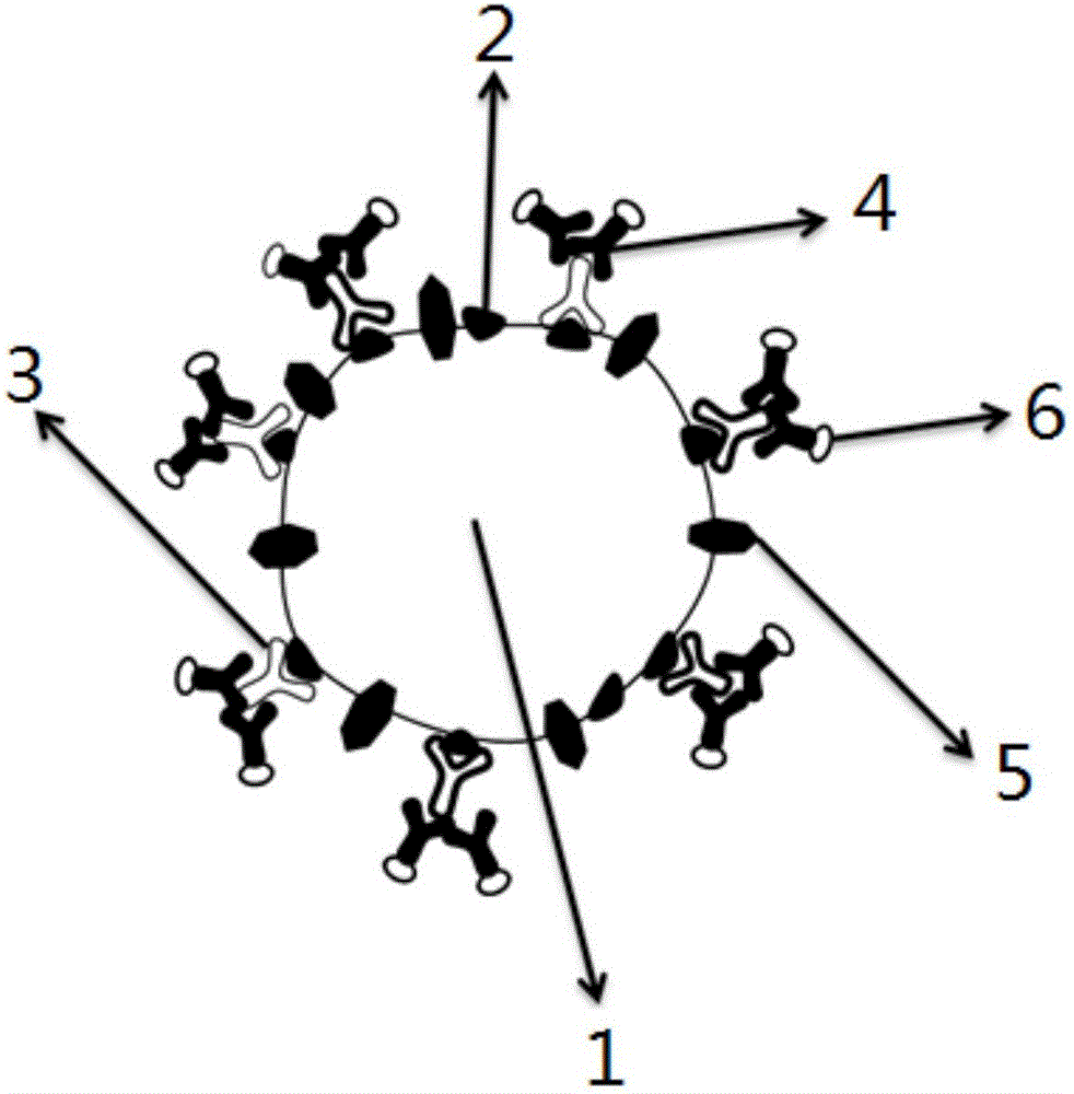
[0023] The PBS buffer involved in this embodiment is pH=7.4, the PBS buffer that concentration is 10mM; The normal cell membrane dye (the third fluorescent molecule) involved is PKH67, and its reaction solution is the Diluent C solution that Sigma company provides; Single-molecule localization super-resolution optical imaging technology is PALM technology, and the imaging buffer involved is a phosphate solution with pH=8.0, which is equipped with 136mM β-mercaptoethanol, 5% glucose, 0.5mg / mL glucose oxidase and 40μg catalase / mL; the tumor cell exosomes involved are breast cancer cell (SKBR3 cells) exosomes, and the normal cells are human embryonic lung fibroblasts (MRC-5 cells); the tumor cell exosome membranes involved The surface receptor is HER2, the primary antibody used in indirect immunofluorescence is rabbit anti-human HER2, and the secondary antibody is goat anti-rabbit IgG l...


PUM


Abstract

A super-resolution optical imaging method based on a single-molecule positioning process is disclosed and used for observing an interaction process between a tumor cell exosome and a normal cell. The method includes (1) extracting the tumor cell exosome by utilizing a kit, (2) respectively labeling a membrane of the tumor cell exosome and a membrane surface receptor of the tumor cell exosome by using a first fluorescent molecule and a second fluorescent molecule, and staining the normal cell by using a third fluorescent molecule, and (3) observing the interaction process between the tumor cell exosome and the normal cell through a super-resolution optical microscope based on the single-molecule positioning process. The method overcomes the problem that exosomes cannot be observed carefully in the prior art due to a diffraction limit, and provides a novel technical means for researching exosome-mediated cancer metastasis mechanisms and for treating and researching cancer metastasis and spreading.

Description

technical field [0001] The invention relates to a super-resolution optical imaging method based on a single-molecule localization method, which is used to observe the interaction process between tumor cell exosomes and normal cells, and belongs to biophotonics technology. Background technique [0002] Exosomes are a type of vesicles secreted by cells, with a size of about 30-100 nm. The outer layer of this interesting nano-scale vesicle is a lipid membrane, on which specific signaling molecules are expressed, while the interior contains biomolecules such as proteins and RNA. Exosomes play a very important role in intercellular communication and material exchange. Existing studies have shown that the interaction between exosomes and recipient cells is mainly divided into three types: 1) exosomes bind to the surface of recipient cells through exosome adhesion molecules on the surface of recipient cells; 2) The exosomes directly fuse with the recipient cells; 3) The exosomes ...

Claims


Application Information

Patent Timeline
IPC IPC(8): G01N21/64G01N33/58G01N33/574G01N33/50
CPCG01N21/6458G01N21/6486G01N33/5032G01N33/574G01N33/582
Inventor 宗慎飞陈晨王著元崔一平
Owner SOUTHEAST UNIV
Learn More