Ultrasound contrast imaging method and apparatus

A technology of contrast-enhanced ultrasound and imaging methods, which is applied in ultrasound/sonic/infrasonic diagnosis, sonic diagnosis, infrasonic diagnosis, etc. It can solve the problems of limited CTR improvement of contrast images, poor resolution of contrast images, and limited penetration of contrast images. Achieve the effect of improving CTR and contrast resolution, increasing dynamic range, and suppressing tissue residue

Active Publication Date: 2016-10-26
SHENZHEN MINDRAY BIO MEDICAL ELECTRONICS CO LTD
View PDF7 Cites 3 Cited by
  • Summary
  • Abstract
  • Description
  • Claims
  • Application Information

AI Technical Summary

Problems solved by technology

[0006] However, because the second harmonic signal attenuates quickly in the tissue, the penetration of the contrast image is limited, and because the tissue also generates the second harmonic component and mixes with the second harmonic component of the contrast agent, the The CTR improvement of contrast images is limited
The high energy required for the generation of the second harmonic signal will damage the microbubbles and reduce the duration of the contrast agent in the human body, and the sub-harmonic will lead to poor contrast image resolution due to its low frequency
Therefore the processing method of above-mentioned two prior art all can not reach desirable effect

Method used


Image

  • Ultrasound contrast imaging method and apparatus
  • Ultrasound contrast imaging method and apparatus
  • Ultrasound contrast imaging method and apparatus

Examples


Embodiment 1

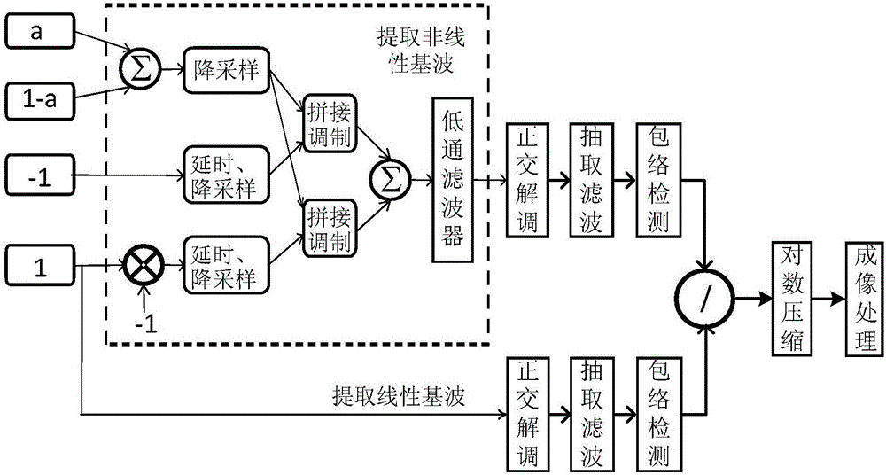
[0046] Such as figure 1 As shown, a contrast-enhanced ultrasound imaging method provided in Embodiment 1 of the present invention specifically includes the following steps:

[0047] S1. Transmit 4 pulse waveform sequences, and obtain the echo signal of each pulse. The four pulses have different amplitude weights; in this embodiment, the amplitude weights of the four pulses are a, -1, 1 and 1-a respectively, where 0

[0048] S2. Process the echo of each pulse, extract the nonlinear fundamental wave, and obtain the amplitude information of the nonlinear fundamental wave;

[0049] S3. Select any one or multiple pulse echoes for processing, extract the linear fundamental wave, and obtain the amplitude information of the linear fundamental wave;

[0050] S4. Using the amplitude information of the linear fundamental wave and the amplitude information of the nonlinear fundamental wave to generate a nonlinear parameter;

[0051] S5. Perform imaging processing on the nonlinear ...

Embodiment 2

[0064] In the present invention, the nonlinear component of the echo is not limited to the nonlinear fundamental wave, and may also be the second harmonic in the echo.

[0065] Such as Figure 8 As shown, a contrast-enhanced ultrasound imaging method provided by an embodiment of the present invention provides a procedure for performing nonlinear parametric imaging using second harmonics.

[0066] Specifically, the embodiment of the present invention includes the following steps:

[0067] S1. Transmit two pulse waveform sequences, and obtain the echo signal of each pulse. The two pulses have different amplitude weights; in this embodiment, the amplitude weights of the two pulses are -1 and 1 respectively;

[0068] S2. Process the echo of each pulse, extract the second harmonic, and obtain the amplitude information of the second harmonic;

[0069] S3. Select one or more echoes of the pulses for processing, extract the linear fundamental wave, and obtain the amplitude informat...

Embodiment 3

[0081] In the present invention, the nonlinear component of the echo can also be the sum of the amplitudes of the nonlinear fundamental wave and the second harmonic.

[0082] A contrast-enhanced ultrasound imaging method provided by an embodiment of the present invention provides a process for performing nonlinear parametric imaging using the sum of the amplitudes of the nonlinear fundamental wave and the second harmonic.

[0083] Specifically, the embodiment of the present invention includes the following steps:

[0084] S1. Transmit 4 pulse waveform sequences, and obtain the echo signal of each pulse. The four pulses have different amplitude weights; in this embodiment, the amplitude weights of the four pulses are a, -1, 1 and 1-a respectively, where 0

[0085] S2. Processing the echoes of each pulse, extracting the nonlinear fundamental wave and the second harmonic respectively, obtaining the amplitude information of the two, and calculating the sum of the amplitude i...


PUM


Abstract

The invention provides an ultrasound contrast imaging method and apparatus. The method comprises: S1. transmitting a plurality of pulse waveform sequences, and acquiring echo signals of various pulses; S2. processing echoes of the various pulses to extract non-linear components of the echoes, and acquiring amplitude information about the non-linear components; S3. selecting echoes of any one or more pulses for processing to extract linear components of the echoes, and acquiring amplitude information about the linear components; S4. using the amplitude information about the non-linear components and that of the linear components to generate a non-linear parameter; and S5. performing imaging processing on the non-linear parameter. In the method, by simultaneously using amplitude information about linear components and non-linear components of an echo to generate a non-linear parameter, and performing imaging after processing this parameter, tissue residues can be effectively inhibited, and the dynamic range of a contrast agent signal is increased, thereby improving the CTR and contrast resolution of a contrast image.

Description

technical field [0001] The invention relates to the field of contrast-enhanced ultrasound imaging, in particular to a method and device for contrast-enhanced ultrasound imaging. Background technique [0002] In the medical ultrasound imaging system, the probe is usually driven by the transmitting circuit to transmit ultrasonic pulses into the human body, and the visible ultrasound of the human tissue is obtained by receiving and processing the echo carrying the characteristic information of the human tissue by using the reflection of the ultrasonic wave at the interface of the human tissue image. However, due to the existence of reverberation and the limitation of resolution, the display of weak borders and small blood vessels in ultrasound images is blurred, and sometimes even cannot be displayed. The contrast agent has a large difference in acoustic impedance from the surrounding tissue, which can change the absorption, reflection, scattering and refraction of sound waves...

Claims


Application Information

Patent Timeline
Patent Type & Authority Applications(China)
IPC IPC(8): A61B8/00G06F19/00
CPCA61B8/00A61B8/13A61B8/481A61B8/5207
Inventor 桑茂栋储霞冒祖华
Owner SHENZHEN MINDRAY BIO MEDICAL ELECTRONICS CO LTD
Why Eureka
Try Eureka