Method for three-dimensional (3D) printing of restoring implant by bony articular surface reconstruction

A 3D printing, bone and joint technology, applied in bone implants, medical science, surgery, etc., can solve problems such as difficult to achieve personalization and precision, achieve personalized anatomical matching, overcome high costs, and save time and cost Effect

Active Publication Date: 2016-11-09
SOUTHERN MEDICAL UNIVERSITY
View PDF9 Cites 32 Cited by
  • Summary
  • Abstract
  • Description
  • Claims
  • Application Information

AI Technical Summary

Problems solved by technology

[0004] The present invention proposes a method for bone articular surface reconstruction by 3D printing and repairing implants, which solves the problem of individuation and precision that is difficult to achieve in traditional intra-articular fracture repair and reconstruction methods, realizes restoration of articular surface height, personalized anatomical shape matching, and manufactures fast and flexible

Method used


Image

  • Method for three-dimensional (3D) printing of restoring implant by bony articular surface reconstruction
  • Method for three-dimensional (3D) printing of restoring implant by bony articular surface reconstruction
  • Method for three-dimensional (3D) printing of restoring implant by bony articular surface reconstruction

Examples


Embodiment 1

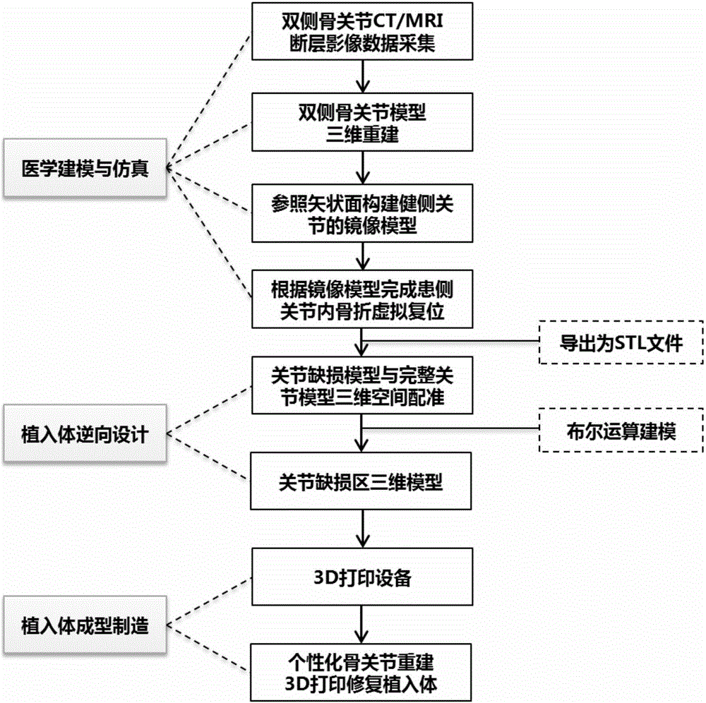
[0032] A method for reconstructing 3D printing and restoring implants of bone articular surface, including the following steps:

[0033] (1) Three-dimensional reconstruction of bone and joint model

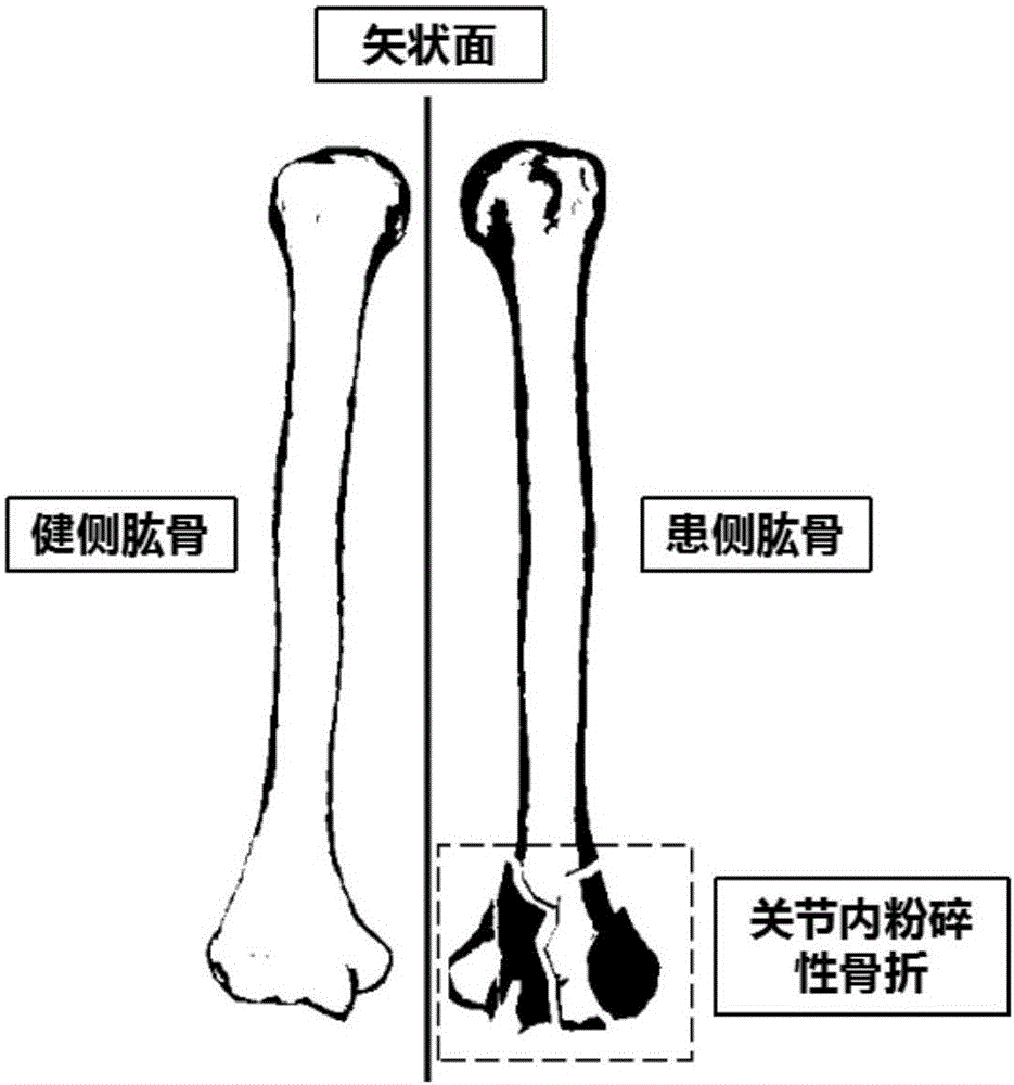
[0034] Collect the computed tomography data of the bilateral limbs of patients with intra-articular fractures, and according to the obtained tomographic Dicom format data, further import the medical three-dimensional reconstruction software to establish the three-dimensional model of the bilateral joints.

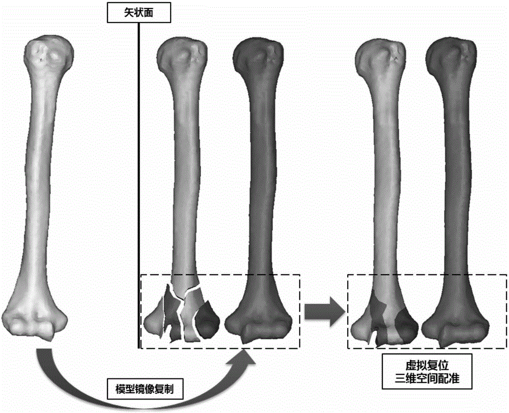
[0035] (2) Virtual reduction and reconstruction of bone and joint defects

[0036] According to the symmetrical morphological characteristics of the human anatomical structure, using the simulation function module of the medical modeling software, the 3D model of the contralateral bone joint established in the above step (1) is mirrored and copied to the affected side in a sagittal symmetrical manner, and the result is The complete structural reference model before the fracture of the ...


PUM


Abstract

The invention provides a method for three-dimensional (3D) printing of a restoring implant by bony articular surface reconstruction. First three-dimensional reconstruction of a bone articular model is performed, and then virtual restoration reconstruction of bony articular reconstruction, reversal design of a bone articular defect area restoring implant and 3D printing forming of a bone articular defect restoring implant are performed. Modeling software is utilized to copy a normal side bone articular three-dimensional model to an affected side in a sagittal plane symmetry mode, so as to obtain a complete structure reference model before the occurrence of fracture of the affected side; 3D printing pro-processing software is utilized to support and generate the model, a corresponding forming material and a quick forming process are selected to perform 3D printing, a support structure of the implant is further removed, and finally an individual bone articular reconstruction restoring implant is obtained. The individual reversal design is performed according to the symmetry morphological structure of the human body, the bony articular surface reconstruction defect restoring implant is quickly formed by combination with a 3D printing technology, not only can a bone articular defect restoring reconstruction be greatly improved, but also the design and manufacturing efficiency of the implant is effectively improved, the cost is reduced, and customized design and individual-based treatment are performed.

Description

Technical field [0001] The invention relates to the technical field of clinical repair and reconstruction of bone and joint injuries and the research and development of implantable medical devices, and in particular to a method for reconstructing 3D printing and repairing implants of bone joint surfaces. Background technique [0002] Accurate articular surface reconstruction is a key factor in the treatment of intra-articular fractures. At present, the clinical classification of fractures mostly uses the internationally accepted AO classification method. The AO classification divides intra-articular fractures into Type B and Type C. Type B is a partial intra-articular fracture and Type C is a complete intra-articular fracture. Both types of intra-articular fractures involve the articular surface of the bone. Based on this The degree of comminution of the articular surface can be further divided into multiple subtypes. Regardless of the classification, for intra-articular fractu...

Claims


Application Information

Patent Timeline
Patent Type & Authority Applications(China)
IPC IPC(8): A61B17/56A61F2/28
CPCA61B17/56A61B2017/564A61F2/28
Inventor 黄文华欧阳汉斌邓羽平谢普生廖政文
Owner SOUTHERN MEDICAL UNIVERSITY
Why Eureka
Try Eureka