Electrochemical biosensor for detecting α-ketoglutarate, preparation method and application thereof

A biosensor, ketoglutaric acid technology, applied in the field of electrochemical detection, can solve the problems of large error, high detection cost, and long time consumption, and achieve the effect of low detection limit, good repeatability, and high sensitivity

Active Publication Date: 2019-02-26
NANJING TECH UNIV
View PDF3 Cites 1 Cited by
  • Summary
  • Abstract
  • Description
  • Claims
  • Application Information

AI Technical Summary

Problems solved by technology

At present, the methods for detecting α-ketoglutarate mainly include colorimetry, high performance liquid chromatography, and fluorescent probes, but these methods have problems such as large errors, long time-consuming, and high detection costs.

Method used


Image

  • Electrochemical biosensor for detecting α-ketoglutarate, preparation method and application thereof
  • Electrochemical biosensor for detecting α-ketoglutarate, preparation method and application thereof
  • Electrochemical biosensor for detecting α-ketoglutarate, preparation method and application thereof

Examples


Embodiment 1

[0024] Embodiment 1 Construction of working electrode

[0025] 1. Preparation of graphene oxide and chloroauric acid mixed electrolyte

[0026] (1) Graphene oxide dispersion: Take 10 mg of graphene oxide powder (purchased from Suzhou Hengqiu Graphene Technology Co., Ltd.), add it to 10 mL of deionized water, crush it with an ultrasonic crusher for 20 minutes, and prepare 1 mg / mL of graphene oxide powder. Graphene dispersion.

[0027] (2) Chlorauric acid solution: Dissolve 412 mg of chloroauric acid in 10 mL of deionized water to prepare a 0.01 M chloroauric acid solution.

[0028] (3) Graphene oxide and chloroauric acid mixed electrolyte: take 200 μL of the chloroauric acid solution prepared in (2) and dissolve it in the graphene oxide dispersion prepared in (1) to obtain a mixed electrolyte.

[0029] 2. Fixed gold nano-graphene composite film

[0030] Polish the glassy carbon electrode with alumina powder with a particle size of 0.3 μm until the electrode surface becomes a...

Embodiment 2

[0036] Example 2 Electrochemical biosensor for detecting α-ketoglutarate

[0037] The electrochemical biosensor used to detect α-ketoglutarate is a classic three-electrode system sensor, including the working electrode, counter electrode and reference electrode prepared in Example 1, the platinum wire electrode as the counter electrode, and the calomel electrode as the counter electrode. Reference electrode.

Embodiment 3

[0038] Example 3 Detection of NADH by an electrochemical biosensor for detecting α-ketoglutarate

[0039] All three electrodes of the sensor in Example 2 were placed in a phosphate buffer solution of pH 7.2 and 0.1 M, and the applied potential was 0.3 volts by chronoamperometry, and 4.5 mM NADH solution was added dropwise to the phosphate buffer solution, every The interval between drops is 100s, and the current-time response curve is obtained, such as Figure 4 . Take the NADH concentration as the abscissa, and the corresponding current as the ordinate, and you can get the scattered points Figure 5 After linear fitting analysis, it can be seen that the linear range of the sensor for NADH detection in Example 2 is 31.9 μM-1.09 mM, and the detection limit is 2 μM, indicating that the sensor has a very high sensitivity.

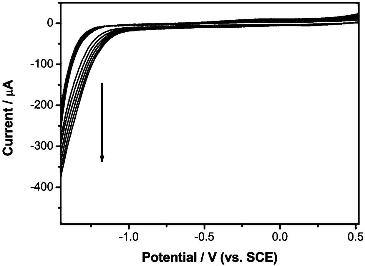
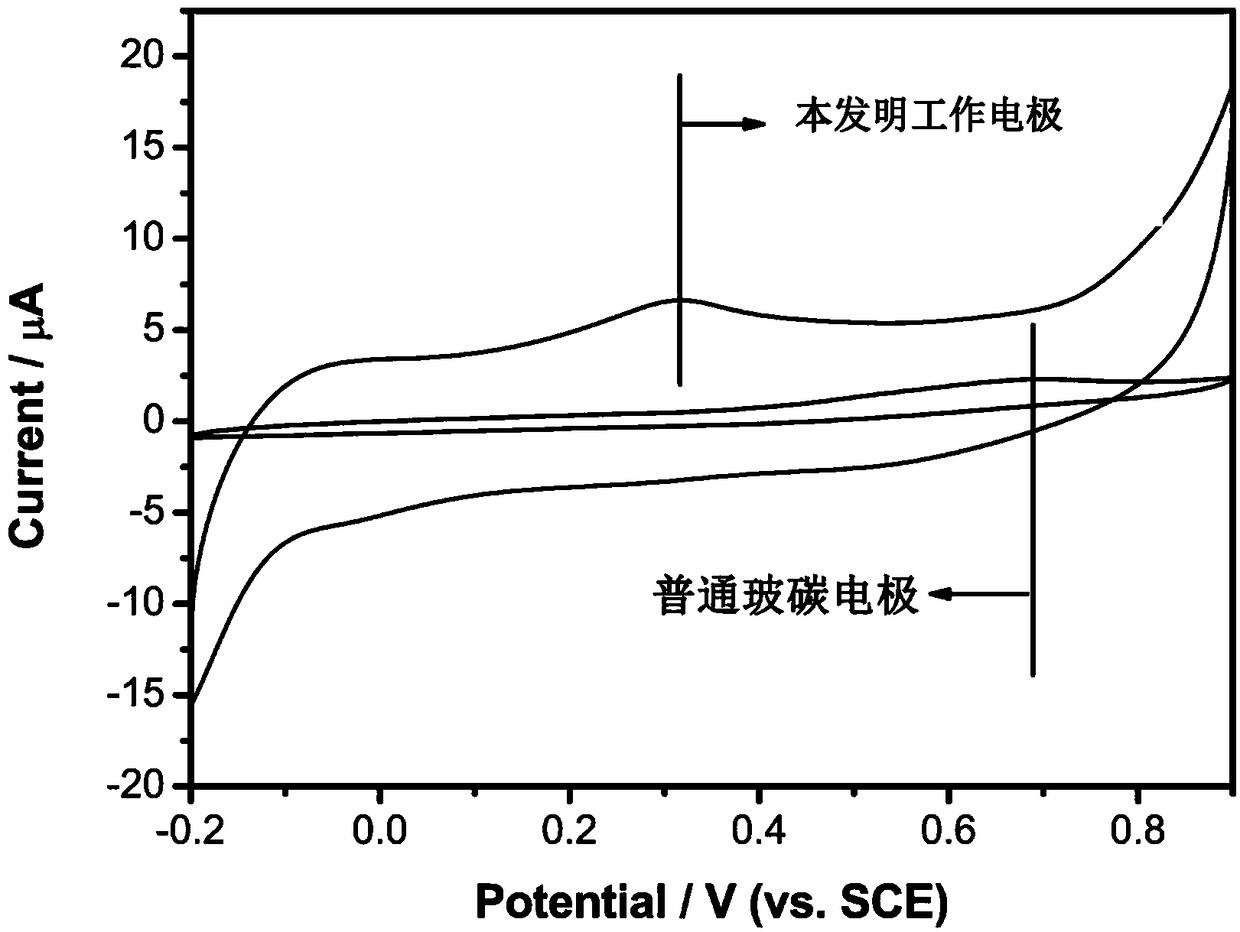
[0040] In addition, compare the cyclic voltammograms of the working electrode prepared in Example 1 (the working electrode of the present invention) and the...


PUM

PropertyMeasurementUnit
concentrationaaaaaaaaaa
volumeaaaaaaaaaa

Abstract

The present invention provides an electrochemical biosensor for detecting alpha-ketoglutarate, a preparation method and applications thereof, and belongs to the field of electrochemical detection. The electrochemical biosensor is a three-electrode system sensor, wherein the working electrode is a glassy carbon electrode with a glutamate dehydrogenase film modified on the surface. The invention further provides the preparation method of the electrochemical biosensor, wherein the preparation method comprises sequentially fixing a nanometer gold-graphene composite film and a glutamate dehydrogenase film on a glassy carbon electrode to prepare the working electrode. According to the present invention, with the application of the sensor to detect the alpha-ketoglutarate, the advantages of high accuracy, short detection time and low cost are provided.

Description

technical field [0001] The invention belongs to the field of electrochemical detection, and in particular relates to an electrochemical biosensor for detecting α-ketoglutarate, a preparation method and an application thereof. Background technique [0002] α-Ketoglutarate is an important metabolic marker in the early detection of tumors, monitoring of microbial industrial fermentation, etc. It is one of the important metabolic intermediates in the tricarboxylic acid cycle and the key to linking intracellular carbon-nitrogen metabolism Nodes are also important precursors for the synthesis of various amino acids and proteins. At present, the methods for detecting α-ketoglutarate mainly include colorimetry, high performance liquid chromatography, and fluorescent probes, but these methods all have problems such as large errors, long time-consuming, and high detection costs. Contents of the invention [0003] Aiming at the shortcomings of the existing detection of α-ketoglutara...

Claims


Application Information

Patent Timeline
Patent Type & Authority Patents(China)
IPC IPC(8): G01N27/26G01N27/327
CPCG01N27/26G01N27/3278
Inventor 黄和彭钢俞亚东徐生盼江凌
Owner NANJING TECH UNIV
Why Eureka
Try Eureka