Amnioscope and preservation method thereof

A preservation method and amnioscopic technology, applied in the field of medical devices, can solve the problems of patients with obvious discomfort symptoms, unsuitable for large-scale promotion and application, complicated operation, etc., to assist the excessive proliferation period, prevent contractures, and maintain anatomical structures Effect

Active Publication Date: 2016-12-14
钛盾生物(南京)有限公司
View PDF3 Cites 8 Cited by
  • Summary
  • Abstract
  • Description
  • Claims
  • Application Information

AI Technical Summary

Problems solved by technology

[0004] At present, a small number of amniotic membrane fixation devices have been used clinically, such as the use of ocular surface biofilm fixation device (BMFD) for sutureless conjunctival sac fresh amniotic membrane coverage, but these devices are not devices with amniotic membrane, and still need to be placed separately during the operation However, the preparation and preservation of amniotic membrane still cannot meet the characteristics of clinical needs at any time, the operation is complicated, and there are obvious discomfort symptoms when patients wear it, so it is not suitable for large-scale promotion and application in my country.

Method used


Image

  • Amnioscope and preservation method thereof
  • Amnioscope and preservation method thereof
  • Amnioscope and preservation method thereof

Examples


Embodiment Construction

[0031] Refer to the attached figure 1 To attach Figure 5 The amnioscope of the present invention and its preservation method are described in detail below.

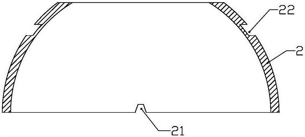
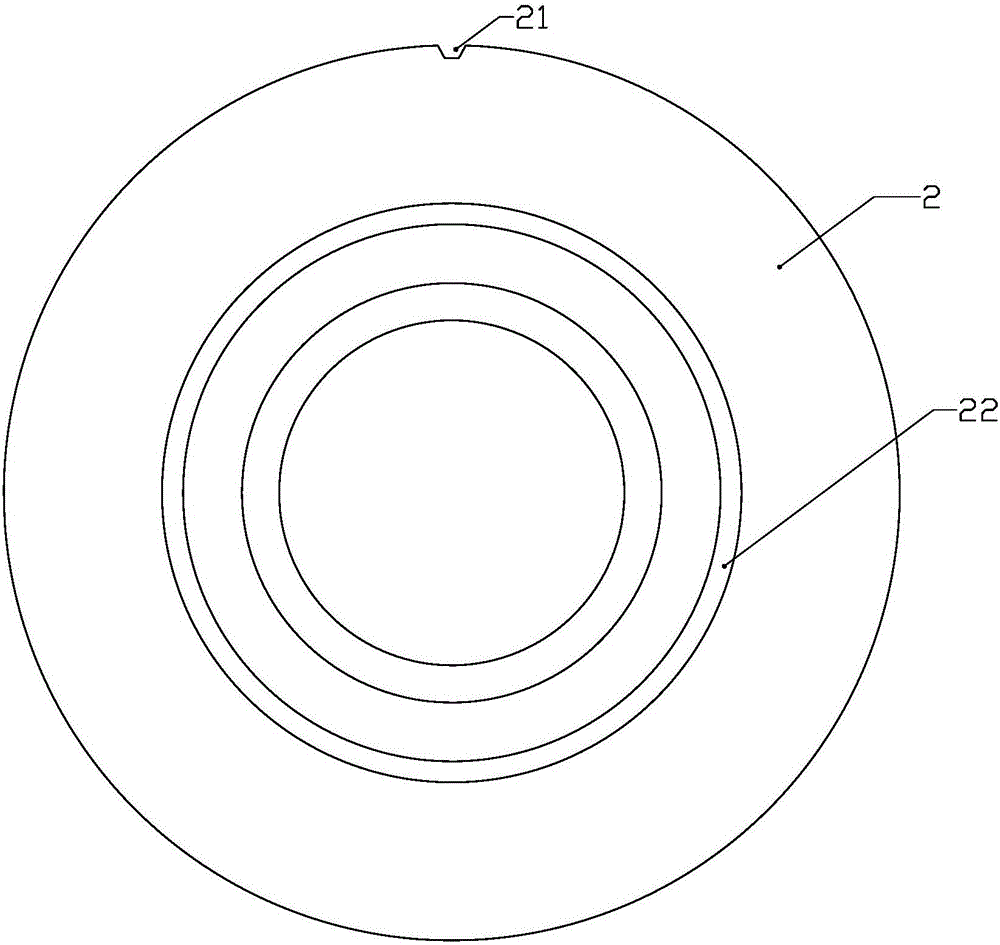
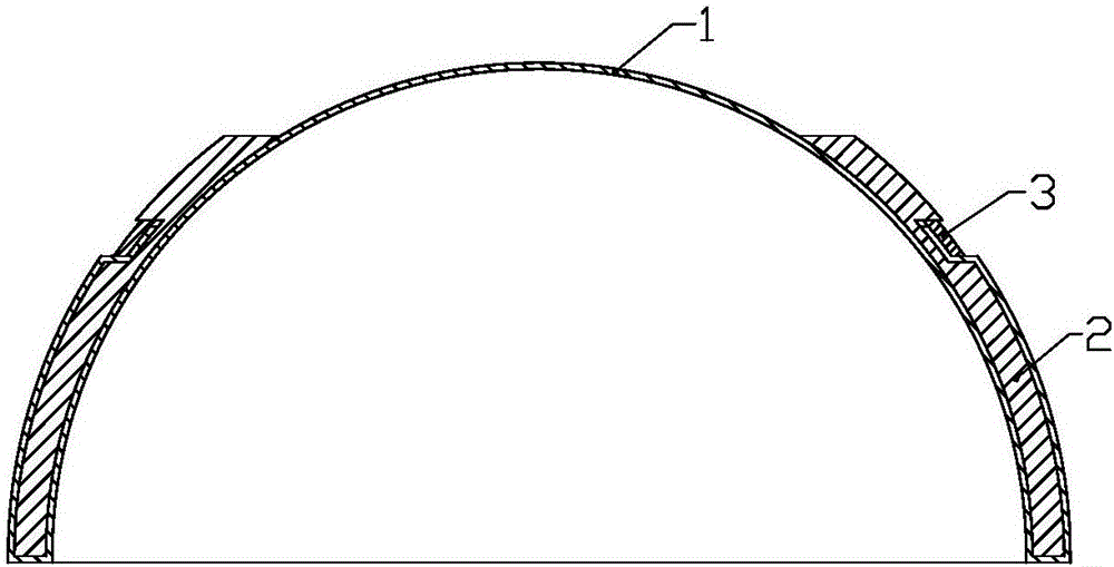
[0032] The structure of the amnioscope of the present invention comprises amniotic membrane 1 and conjunctival sac support device 2, and described conjunctival sac support device 2 is a ring structure whose diameter gradually increases from top to bottom, which is consistent with the curvature of the ocular surface, and adopts polyacrylic It is made of methyl acrylate (PMMA) material and has good tissue compatibility. A trapezoidal groove 21 is opened at the position where the lower part of the conjunctival sac supporting device 2 is in contact with the subocular cavity, so as to drain the subamniotic accumulation Liquid, the upper part of the amniotic membrane 1 is attached to the inner cavity of the conjunctival sac support device 2, and the lower part protrudes from the inner cavity of the conjunctival sac support de...


PUM


Abstract

The invention discloses an amnioscope and a preservation method thereof and belongs to medical instruments. The amnioscope structurally comprises an amniotic membrane and a conjunctival sac supporting device, the conjunctival sac supporting device is of an annular structure with diameter gradually increased from top to bottom and is identical with an ocular surface in radian, a trapezoidal groove is formed in a position where the lower portion of the conjunctival sac supporting device contacts with a lower hole of the ocular surface, and the upper portion of the amniotic membrane is fitted with an inner cavity of the conjunctival sac supporting device while the lower portion of the same extends out of the inner cavity of the same and is fixed to the outer wall of the conjunctival sac supporting device. Compared with the prior art, the amnioscope and the preservation method have the advantages that the amniotic membrane and the conjunctival sac supporting device are integrally arranged, and the amnioscope is preserved in a specific preservation solution and put in certain low-temperature state, so that tissue activity of the amniotic membrane is maintained; the amniotic membrane can be taken for use at any time and is simple to operate, so that uncomfortable symptoms of a patient when wearing the amnioscope are avoided, and the amnioscope has high popularization and application value.

Description

technical field [0001] The invention relates to a medical device, in particular to an amniotic mirror and a preservation method thereof. Background technique [0002] The ocular surface includes conjunctival epithelium, corneal epithelium and tear film. Clinically, ocular surface disease (OSD) mainly refers to lesions of the cornea and conjunctiva, also known as ocular surface tear disease. Severe ocular surface diseases can cause severe vision loss or loss, and ocular surface reconstruction surgery aims to restore visual function. For example, in the early stage of ocular burns, especially severe alkali burns on the ocular surface, if effective treatment cannot be implemented in the early stage, local activation of collagenase and neutral vector enzymes will lead to progressive dissolution of ocular surface tissue and even corneal perforation. Amniotic membrane transplantation can be effectively used to reconstruct the surface of cornea and conjunctiva, not only can be us...

Claims


Application Information

Patent Timeline
Patent Type & Authority Applications(China)
IPC IPC(8): A61F9/00A01N1/02
CPCA01N1/021A61F9/0017A61F2210/00A61F2220/0025A61F2230/0063
Inventor 张晨明
Owner 钛盾生物(南京)有限公司
Why Eureka
Try Eureka