Belly image reconstruction device

An image reconstruction, abdominal technology, applied in the field of image processing, can solve the problems of image loss and underutilization of data, etc.

Inactive Publication Date: 2016-12-14
PEKING UNIV
View PDF2 Cites 5 Cited by
  • Summary
  • Abstract
  • Description
  • Claims
  • Application Information

AI Technical Summary

Problems solved by technology

Therefore, the data is not fully utilized, and the reconstructed image has partial loss

Method used


Image

  • Belly image reconstruction device
  • Belly image reconstruction device
  • Belly image reconstruction device

Examples


Embodiment 1

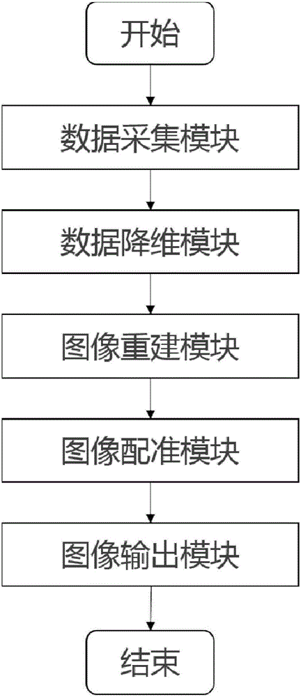
[0072] Embodiment 1, concrete processing steps are as follows:

[0073] 1. The data acquisition module, which collects data on the subject's liver, adopts Golden-Angle Radial subsampling magnetic resonance 3D sequences.

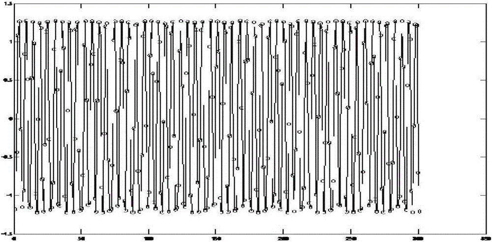
[0074] 2. The data dimensionality reduction module uses the Locally linear embedding (LLE) algorithm to reduce the K-space center data to a one-dimensional sequence.

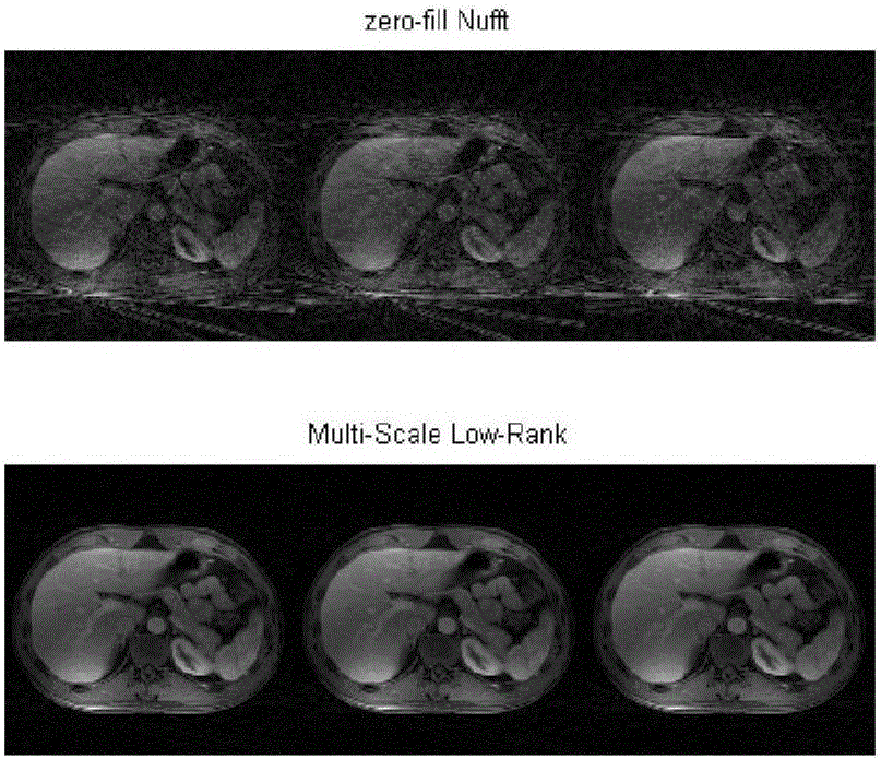
[0075] 3. The image reconstruction module first divides the collected data into 3 categories according to the one-dimensional sequence and the size of the value. For each type of data, the multi-scale low-rank restoration theory is used to reconstruct, and three reconstructed images are obtained.

[0076] 4. The image registration module uses the Lucas-Kanade algorithm to register the three reconstructed images, that is, to register the last two images to the first image.

[0077] 5. The image output module averages the registered 3 images, and finally outputs 1 image.

[0078] Simulation re...

Embodiment 2

[0080] Embodiment 2, concrete processing steps are as follows:

[0081] 1. The data acquisition module, which collects data on the subject’s kidneys, adopts Golden-Angle Radial subsampling magnetic resonance 3D sequences.

[0082] 2. The data dimensionality reduction module uses the Locally linear embedding (LLE) algorithm to reduce the K-space center data to a one-dimensional sequence.

[0083] 3. The image reconstruction module first divides the collected data into 3 categories according to the one-dimensional sequence and the size of the value. For each type of data, the multi-scale low-rank restoration theory is used to reconstruct, and three reconstructed images are obtained.

[0084] 4. The image registration module uses the Lucas-Kanade algorithm to register the three reconstructed images, that is, to register the last two images to the first image.

[0085] 5. The image output module averages the registered 3 images, and finally outputs 1 image.


PUM


Abstract

The present invention provides a belly image reconstruction device. The device is reconstructed in the free breathing, a Golden-Angle Radial 3D rapid scanning sequence is employed to obtain a liver image. The locally linear embedding (LLE) in the manifold learning method is employed to perform the dimension reduction process of the K space center data, and a respiratory curve is obtained; the data packet is performed according to the respiratory curve, and each group of data is subjected to reconstruction through adoption of a multi-scale low-order recovery theory; and finally, each group of reconstructed data is subjected to registering so as to eliminate the motion artifact and obtain a final image. The belly image reconstruction device does not reject any data with no need for an operator's manual participation, and is high in degree of automation and low in application cost, etc.

Description

technical field [0001] The invention relates to a device for abdominal image reconstruction, which belongs to the technical field of image processing. Background technique [0002] Currently, magnetic resonance imaging plays a vital role in the clinical diagnosis and treatment of abdominal lesions. However, MRI takes a long time, and the abdomen will be affected by breathing movements. Respiratory movement will cause movement and deformation of organs, which will lead to respiratory movement artifacts in the image, which will reduce the resolution and signal-to-noise ratio of the image. In the process of image-guided interventional therapy, there will be inconsistencies between the static guidance information and the position of the moving structure. The phenomenon. [0003] At present, the commonly used methods for suppressing respiratory motion artifacts in abdominal images are breath-holding method and respiratory gating method. The breath-hold method is to acquire ima...

Claims


Application Information

Patent Timeline
Patent Type & Authority Applications(China)
IPC IPC(8): G06T11/00
CPCG06T11/005
Inventor 吕骏张晓东王霄英张珏方竞
Owner PEKING UNIV
Why Eureka
Try Eureka