
Method for separating exosomes from serum by using immunomagnetic beads

A technology of immune magnetic beads and exosomes, applied in the field of biomedicine, can solve the problems of high cost, short time-consuming exosomes, time-consuming and labor-intensive problems

Inactive Publication Date: 2017-01-04
EAST CHINA UNIV OF SCI & TECH
View PDF3 Cites 25 Cited by
  • Summary
  • Abstract
  • Description
  • Claims
  • Application Information

AI Technical Summary

Problems solved by technology

The centrifugation method has a high yield of exosomes, but it is time-consuming and labor-intensive, and is not suitable for a large number of samples
The purity of exosomes extracted by chromatography is high, but its separation is easily interfered by foreign proteins, and the operation stability is poor
It takes a short time to precipitate exosomes with macromolecular polymers, but the disadvantage is that the purity of exosomes obtained is low
The above-mentioned techniques can only isolate the total exosomes in the serum, and lack the specific marker exosomes (such as CD9 + / CD63 + Exosomes), the immunomagnetic bead kit method can separate exosomes with specific markers, but this technology is only mastered by a few international giants such as SBI in the United States and MBL in Japan, and the cost of use is high

Method used


Image

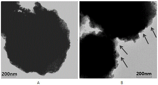
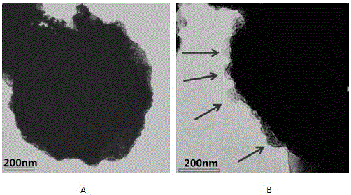
  • Method for separating exosomes from serum by using immunomagnetic beads
  • Method for separating exosomes from serum by using immunomagnetic beads

Examples


Embodiment 1

[0022] Example 1. Immunomagnetic Bead Separation of CD9 in Serum + Exosomes

[0023] one. method

[0024] (1) Collect fresh serum, put it in a centrifuge, and centrifuge at 2000g at room temperature for 30 minutes, discard the cell debris and precipitate, and take the centrifuged supernatant (if the serum is in a frozen state, it needs to be thawed in a water bath at 26°C before centrifuging).

[0025] (2) Vortex streptavidin-modified Dynabeads® MyOne™ Streptavidin T1 magnetic beads. After the magnetic bead suspension is uniform, take 5 microliters of the magnetic bead suspension and add it to 500 microliters of separation buffer, suspend and wash, and magnetically Magnetic beads were separated by rack and washed three times. After washing is complete, resuspend the beads in 500 µl of separation buffer.

[0026] (3) Take 5 microliters of the mouse anti-human biotinylated CD9 monoclonal antibody produced by Ancell Company and add it to the magnetic bead suspension obtained ...

Embodiment 2

[0030] Example 2. Immunomagnetic Bead Separation of CD63 in Serum + Exosomes

[0031] one. method

[0032] (1) Collect fresh serum, put it in a centrifuge, and centrifuge at 2000g at room temperature for 30 minutes, discard the cell debris and precipitate, and take the centrifuged supernatant (if the serum is in a frozen state, it needs to be thawed in a water bath at 26°C before centrifuging).

[0033] (2) Vortex streptavidin-modified Dynabeads® MyOne™ Streptavidin T1 magnetic beads. After the magnetic bead suspension is uniform, take 5 microliters of the magnetic bead suspension and add it to 500 microliters of separation buffer, suspend and wash, and magnetically Magnetic beads were separated by rack and washed three times. After washing is complete, resuspend the beads in 500 µl of separation buffer.

[0034] (3) Take 5 microliters of the mouse anti-human biotinylated CD63 monoclonal antibody produced by Ancell Company and add it to the magnetic bead suspension obtaine...


PUM

PropertyMeasurementUnit
Diameteraaaaaaaaaa

Abstract

The invention discloses a method for separating exosomes from serum by using immunomagnetic beads. The method comprises the following steps: pretreating the serum, coupling a monoclonal antibody with magnetic beads, and separating the exosomes from the serum by using an antibody-magnetic bead compound. The method has the advantages as follows: the method is easy to operate and low in cost; by the method, the exosomes containing specific markers can be separated from the serum.

Description

technical field [0001] The invention relates to the field of biomedicine, in particular to a method for separating exosomes in serum by using immunomagnetic beads. Background technique [0002] Immunomagnetic bead separation technology is an immunological technology developed based on antigen-antibody binding. It mainly relies on modifying amino groups, carboxyl groups, streptavidin and other groups on the surface of magnetic beads. These groups can be used for covalent or non-covalent coupling with antibodies, which can be used to bind corresponding antigens. It can achieve the separation of corresponding antigenic substances, and is widely used in nucleic acid extraction, separation of membrane structure substances such as cell separation. [0003] Exosomes (exosomes) are a type of membrane-structured vesicles with a diameter of 30-150 nm, which are widely found in blood, urine, saliva and other body fluids. The surface of the exosome membrane contains CD9, CD63 and CD81...

Claims


Application Information

Patent Timeline
IPC IPC(8): G01N1/34G01N1/40
CPCG01N1/34G01N1/405
Inventor 叶邦策倪自鹏尹斌成
Owner EAST CHINA UNIV OF SCI & TECH
Learn More