Whole slide digital pathological image processing and analysis method

A digital pathology and image processing technology, applied in the field of medical digital image processing, can solve the problems of lack of objective quantitative data, limited changes, and time-consuming full-section image analysis, to improve accuracy and analysis speed, improve accuracy and The effect of efficiency

Inactive Publication Date: 2017-02-15
诸暨微因生物科技有限公司
View PDF6 Cites 11 Cited by
  • Summary
  • Abstract
  • Description
  • Claims
  • Application Information

AI Technical Summary

Problems solved by technology

[0003] Although digital scanning slices and digital slice image analysis technologies are gradually being promoted and applied in major hospitals and research institutions, doctors mainly browse digital pathological images of full slices with the naked eye, and analyze and diagnose them based on experience. There is no objective quantitative data, and subjectivity Strong, analysis and diagnosis based on personal experience, different pathologists have different judgments on the same slice; and the changes that can be judged by the naked eye are limited, it is difficult to make a complete observation of the pathological phenomena suggested in the tissue slice
[0004] Although the prior art also uses computers to analyze digital pathological images of whole slices, it usually only selects and analyzes local images of multiple fields of view, and these pre-selected areas are considered to represent biologically important expressions. However, , the region assignment is subjective and depends on the experience of the observer, once the selection bias will lead to the lack of reproducibility of the results
Furthermore, due to the high-throughput characteristics of full-slice digital scan images, usually the size of a full-slice digital scan image is between hundreds of MB or even several GB. Therefore, the analysis of full-slice images in the prior art is time-consuming. long, failed to gain popularity
At the same time, the analysis of the whole slice image in the prior art is only based on the analysis of pixels, and the analysis result is simply to give the positive percentage according to the pixels.
Pixel-based WSI analysis can be done very simply and directly, because adjacent regions are simply merged together without considering the object size and the diversity of boundary connections, resulting in inaccurate analysis results

Method used


Image

  • Whole slide digital pathological image processing and analysis method
  • Whole slide digital pathological image processing and analysis method
  • Whole slide digital pathological image processing and analysis method

Examples


Embodiment 1

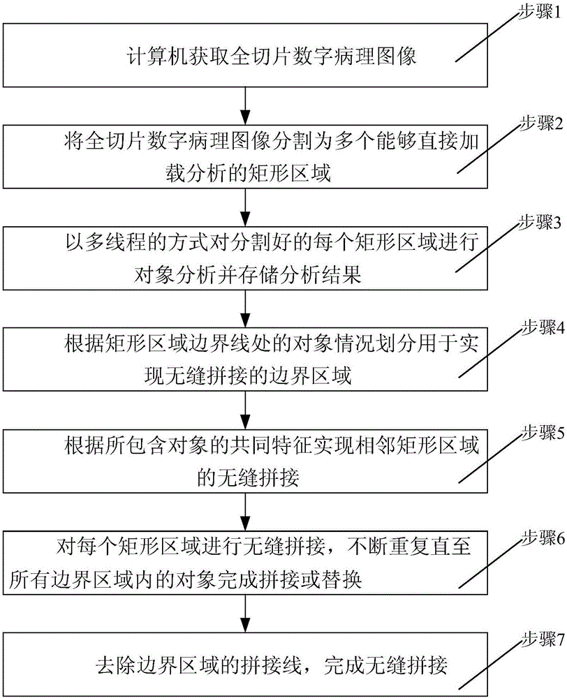
[0056] Finding the tumor area requires the use of artificial intelligence in the software function. In this example, the tissue in the whole section is divided into two categories, tumor tissue and normal tissue, and the object of study is the quantification and grading of nuclei in the tumor region. Through this analysis, combined with the Figure 7-10 , to specifically describe the embodiment of the present invention, the analysis step includes three parts: image segmentation and analysis, seamless splicing and data export.

[0057] 1. Image Segmentation and Analysis

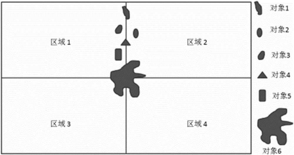
[0058] According to the set protocol file, the tissue is searched and classified. The tumor tissue part is composed of small pieces, which can be analyzed directly. Only one bottom tumor area is too large to be loaded in one frame, so it is divided into about 20 pieces. sub-regions such as Figure 7 shown.

[0059] 2. Seamless splicing

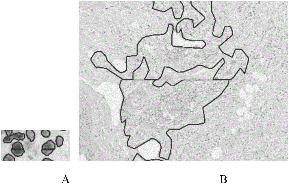
[0060] 1) Figure 7 As shown, there are gaps inside the spliced ​​t...


PUM


Abstract

The invention discloses a whole slide digital pathological image processing and analysis method, and belongs to the field of the whole slide digital pathological image processing and analysis optimization method. The method comprises the steps that step one, a whole slide image (WSI) is segmented into multiple areas, and each area is analyzed; and step two, objects near the boundary of the areas are spliced. The disadvantages of the existing histopathological analysis software can only browse the whole slide digital image and cannot realize analysis or seamless splicing of the whole slide image and is low in efficiency, long in time consumption and blurred in image can be overcome. The main purpose of the method is to process whole slide digital pathological image analysis, segment the whole slide image into multiple areas with the help of the computer and the fine algorithm and automatically perform high-precision multi-vision seamless splicing and processing so that the analysis time can be shortened and the analysis precision can be enhanced, and the high-quality visual data can be acquired to be applied to various fields of pathological histology.

Description

technical field [0001] The invention belongs to the field of medical digital image processing, and in particular relates to a method for processing and analyzing full slice digital pathological images. Background technique [0002] At present, pathological laboratory medicine is developing towards the "five modernizations", namely standardization, automation, informationization, humanization and clinicalization. The emergence of high-definition, high-resolution digital scanning slices has completed the conversion of optical microscopic images to digital microscopic images, but this is only the basis of digital pathology technology. Research and development of digital pathology image processing and analysis methods have a significant impact on digital pathology technology. development plays a more decisive role. [0003] Although digital scanning slices and digital slice image analysis technologies are gradually being promoted and applied in major hospitals and research inst...

Claims


Application Information

Patent Timeline
Patent Type & Authority Applications(China)
IPC IPC(8): G06T7/11G06T7/187
CPCG06T2207/10008G06T2207/30004
Inventor 阮俞敏子阮建忠陈韶炜
Owner 诸暨微因生物科技有限公司
Why Eureka
Try Eureka