Supercharge Your Innovation With Domain-Expert AI Agents!

Sparkle artifact detection in Ultrasound color flow

A technology of color and artifacts, applied in the re-radiation of sound waves, blood flow measurement devices, ultrasonic/sonic/infrasonic diagnosis, etc., can solve the problem of loss of sensitivity

Active Publication Date: 2017-03-22
SIEMENS MEDICAL SOLUTIONS USA INC
View PDF9 Cites 2 Cited by
  • Summary
  • Abstract
  • Description
  • Claims
  • Application Information

AI Technical Summary

Problems solved by technology

Typically, the gain or transmit power is reduced to reduce this flicker artifact, but at the expense of loss of sensitivity
Flashes can be identified using spatial variation of the color flow, but may reduce aliased flow or perturbation and artifacts

Method used


Image

  • Sparkle artifact detection in Ultrasound color flow
  • Sparkle artifact detection in Ultrasound color flow
  • Sparkle artifact detection in Ultrasound color flow

Examples


Embodiment Construction

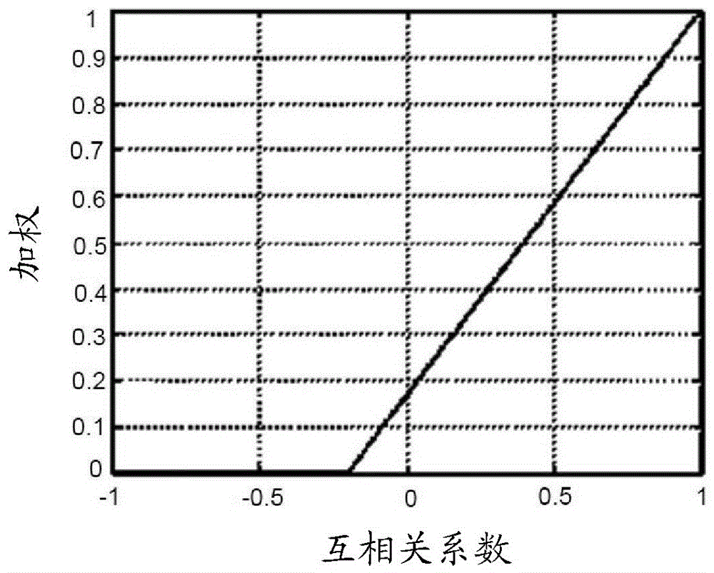
[0013] Detecting Glitter Artifacts in Ultrasound Imaging. Flashes, sometimes called flickers, are more random than fluid or tissue motion. Varying the pulse repetition frequency (PRF) can yield different information for flash artifacts. Velocity estimated from blood flow is generally independent of PRF. By generating two images with different PRFs and performing a normalized cross-correlation, a weighting matrix can be applied to produce images with reduced flash (e.g. flow only or clearer flow) or with reduced flow (e.g. artifacts only or enhanced stones) image. Flash artifacts are detected without compromising sensitivity by using PRF changes.

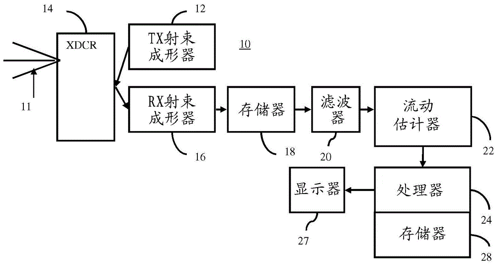
[0014] figure 1 One embodiment of a method for glint artifact detection in color flow is shown. Color flow is used to indicate spatial motion imaging, such as fluid or tissue motion. "Color" is used to distinguish from spectral Doppler imaging, where the power spectrum is estimated for range gating. Colored "flow" data may be ...


PUM


Abstract

Sparkle in color flow imaging is detected. Color flow data is estimated with different pulse repetition frequency (PRF). By correlating the color flow data estimated with different PRFs, sparkle is identified. Color flow images may be filtered to reduce motion while maintaining the sparkle region (e.g., kidney stone imaging) or reduce the sparkle region while maintaining motion (e.g., remove sparkle as system noise).

Description

Background technique [0001] This present embodiment relates to glint detection in ultrasound color flow imaging. Color flow imaging is susceptible to artifacts when there is a loss of correlation in the backscattered echoes. Due to surface roughness, loss of correlation can be indicative of underlying physiology, such as kidney stones. Correlation loss may alternatively be due to system imperfections, such as phase noise or reverberation noise. Typically, gain or transmit power is reduced to reduce this flicker artifact, but at the expense of loss of sensitivity. Using the spatial variation of the color flow can identify glints, but possibly reduce aliased flows or disturbances and artifacts. Contents of the invention [0002] By way of introduction, the preferred embodiments described below include methods, systems, computer readable media, and instructions for glint detection in color flow imaging. Estimate color flow data using different pulse repetition frequencies (...

Claims


Application Information

Patent Timeline
Patent Type & Authority Applications(China)
IPC IPC(8): G06K9/62G06K9/40
CPCG06V10/30G06V2201/031G06F18/22G01S7/5202G01S7/52026G01S7/5205G01S15/8988A61B8/5269G01S7/52071A61B8/06A61B8/463A61B8/5223A61B8/488A61B8/5207G06T7/254G06T2207/10132G06T5/70
Inventor C.H.徐王景源
Owner SIEMENS MEDICAL SOLUTIONS USA INC
Features
  • R&D
  • Intellectual Property
  • Life Sciences
  • Materials
  • Tech Scout
  • Unparalleled Data Quality
  • Higher Quality Content
  • 60% Fewer Hallucinations
Learn More