Imaging method and device based on plane waves

An imaging method and plane wave technology, applied in blood flow measurement devices, acoustic wave diagnosis, infrasound wave diagnosis, etc., can solve the problem of not being able to quantitatively reflect blood flow information in a specific area, and achieve the effect of simplifying the operation process and improving efficiency

Active Publication Date: 2017-05-31
VINNO TECH (SUZHOU) CO LTD
View PDF5 Cites 14 Cited by
  • Summary
  • Abstract
  • Description
  • Claims
  • Application Information

AI Technical Summary

Problems solved by technology

[0006] Embodiments of the present invention provide a plane wave-based imaging method and device to solve the technical problem i

Method used


Image

  • Imaging method and device based on plane waves
  • Imaging method and device based on plane waves
  • Imaging method and device based on plane waves

Examples


Embodiment 1

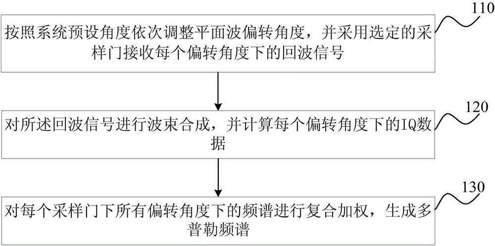
[0045] figure 1 It is a schematic flow chart of the plane wave-based imaging method provided by Embodiment 1 of the present invention. This embodiment is applicable to the situation of reducing the power consumption of the beamformer. The method can be performed by a device for reducing the power consumption of the beamformer. The device can be It is implemented in a software / hardware manner, and can be integrated into ultrasonic imaging equipment, especially portable and hand-held ultrasonic imaging equipment.

[0046] see figure 1 , the method for reducing the power consumption of a beamformer, comprising:

[0047] S110, sequentially adjusting the deflection angle of the plane wave according to the system preset angle, and using a selected sampling gate to receive echo signals at each deflection angle.

[0048]N plane wave angles A1, A2, ..., AN preset in the internal file of the ultrasound imaging system can be read, such as -15°, -10°, 0°, 10°, 15°. The number N of plan...

Embodiment 2

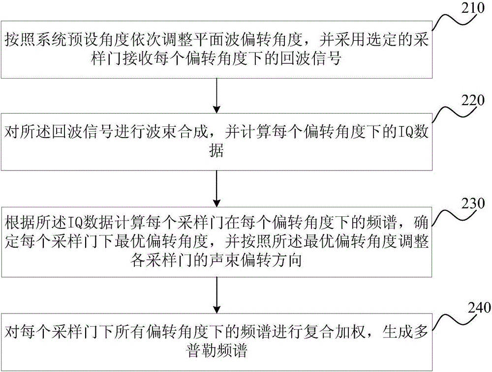
[0056] figure 2 It is a schematic flowchart of the plane wave-based imaging method provided by Embodiment 2 of the present invention. This embodiment is optimized on the basis of the above-mentioned embodiments. In this embodiment, after calculating the IQ data at each deflection angle, the following steps are added: according to the IQ data, calculate the Spectrum of , determine the optimal deflection angle under each sampling gate.

[0057] see figure 2 , an imaging method based on plane waves, including:

[0058] S210, sequentially adjusting the deflection angles of the plane wave according to the system preset angles, and using a selected sampling gate to receive echo signals at each deflection angle.

[0059] S220. Perform beamforming on the echo signals, and calculate IQ data at each deflection angle.

[0060] S230. Calculate the frequency spectrum of each sampling gate at each deflection angle according to the IQ data, determine the optimal deflection angle for ea...

Embodiment 3

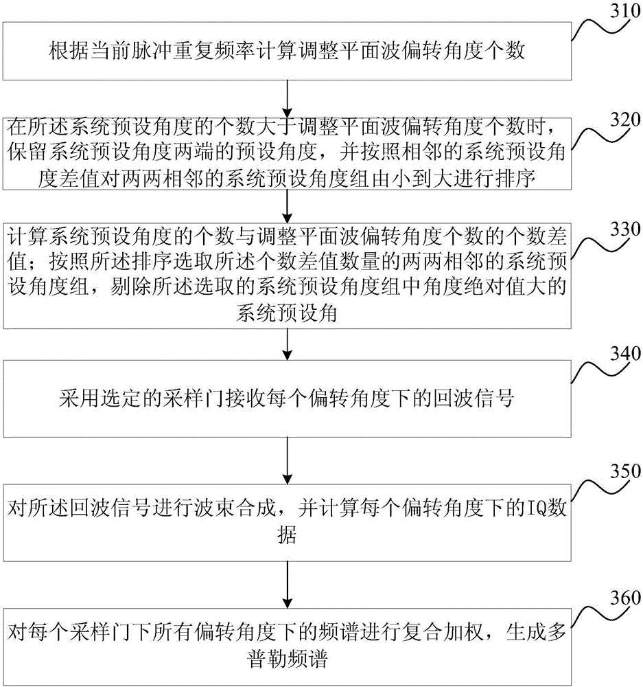
[0068] image 3 It is a schematic flowchart of the plane wave-based imaging method provided by Embodiment 3 of the present invention. This embodiment is optimized on the basis of the above-mentioned embodiments. In this embodiment, adjusting the plane wave deflection angles in sequence according to the system preset angles includes: calculating and adjusting the number of plane wave deflection angles according to the current pulse repetition frequency; When the number of system preset angles is greater than the number of adjusted plane wave deflection angles, the preset angles at both ends of the system preset angles are retained, and the pairwise adjacent system preset angle groups are divided by Sort from small to large; calculate the difference between the number of system preset angles and the number of adjusted plane wave deflection angles, and select two adjacent system preset angle groups according to the sorting ; Eliminate system default angles with larger absolute v...


PUM


Abstract

The invention discloses an imaging method and device based on plane waves. The imaging method includes the steps that deflection angles of the plane waves are sequentially adjusted according to a preset angle of a system, and echo signals of all the deflection angles are received by selected sampling gates; the echo signals are subjected to beam forming, and IQ data of all the deflection angles is calculated; frequency spectrums of all the deflection angles of all the sampling gates are subjected to composite weighting, and a Doppler frequency spectrum is generated. The best deviation angle can be automatically obtained, and automatic deflection of spectral Doppler angles is achieved; instant blood flow velocity information of a plurality of areas can be simultaneously and quantitatively analyzed, the scanning operation procedure of the blood flow velocity is simplified, and the scanning efficiency of the blood blow velocity is improved.

Description

technical field [0001] The invention relates to the technical field of medical ultrasonic imaging, in particular to an imaging method and device based on plane waves. Background technique [0002] Pulse Spectrum Doppler Imaging Technology and Color Doppler Imaging Technology are currently the two most important means in the field of blood flow imaging. Color Doppler imaging technology can provide intuitive blood flow distribution, but lacks the ability to quantitatively analyze blood flow velocity; spectral Doppler imaging technology can accurately and quantitatively detect blood flow velocity, so spectral Doppler imaging technology It is widely used in blood flow detection. [0003] Spectral Doppler imaging relies on the Doppler effect. When the direction of the ultrasound beam is perpendicular to the direction of the blood flow, according to the Doppler effect, the Doppler frequency deviation is almost zero at this time. Therefore, in this case, the sensitivity to blood...

Claims


Application Information

Patent Timeline
IPC IPC(8): A61B8/00A61B8/06
CPCA61B8/00A61B8/06A61B8/4411A61B8/488A61B8/5207
Inventor 吴方刚凌涛
Owner VINNO TECH (SUZHOU) CO LTD
Why Eureka
Try Eureka