Endoscope suture instrument

A suturing device and suture needle technology, applied in the field of endoscopic suturing device, can solve the problems of occupying space, weight, poor work continuity, etc., and achieve the effects of reasonable structure, convenient operation and good operation continuity

Active Publication Date: 2017-05-31
中国人民解放军联勤保障部队第九00医院 +1
View PDF7 Cites 2 Cited by
  • Summary
  • Abstract
  • Description
  • Claims
  • Application Information

AI Technical Summary

Problems solved by technology

The biggest disadvantage of this invention is that it uses a metal needle instead of a thread. The size of the circle of the needle and the amount of tissue it pricks may affect the firmness and sealing of the closure. If the circle is large or contains less tissue, it will still The closure is not tight. Once the fistula still exists, all previous efforts will be for naught. Another disadvantage is that when the suture is wrong, there is no special recovery device, so it is difficult to recover. If it is taken by force, it will aggravate further damage to the tissue and may cause massive bleeding. Do not take it. Because it is a metal object, it takes up space and affects the suture of the next stitch, and the work continuity is poor

Method used


Image

  • Endoscope suture instrument
  • Endoscope suture instrument
  • Endoscope suture instrument

Examples


Embodiment 1

[0052] Please refer to Figure 1 to Figure 4 , Embodiment 1 of the present invention is:

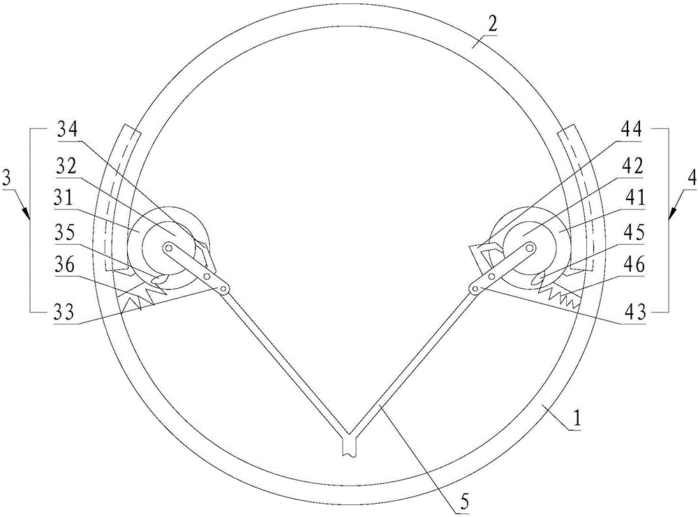
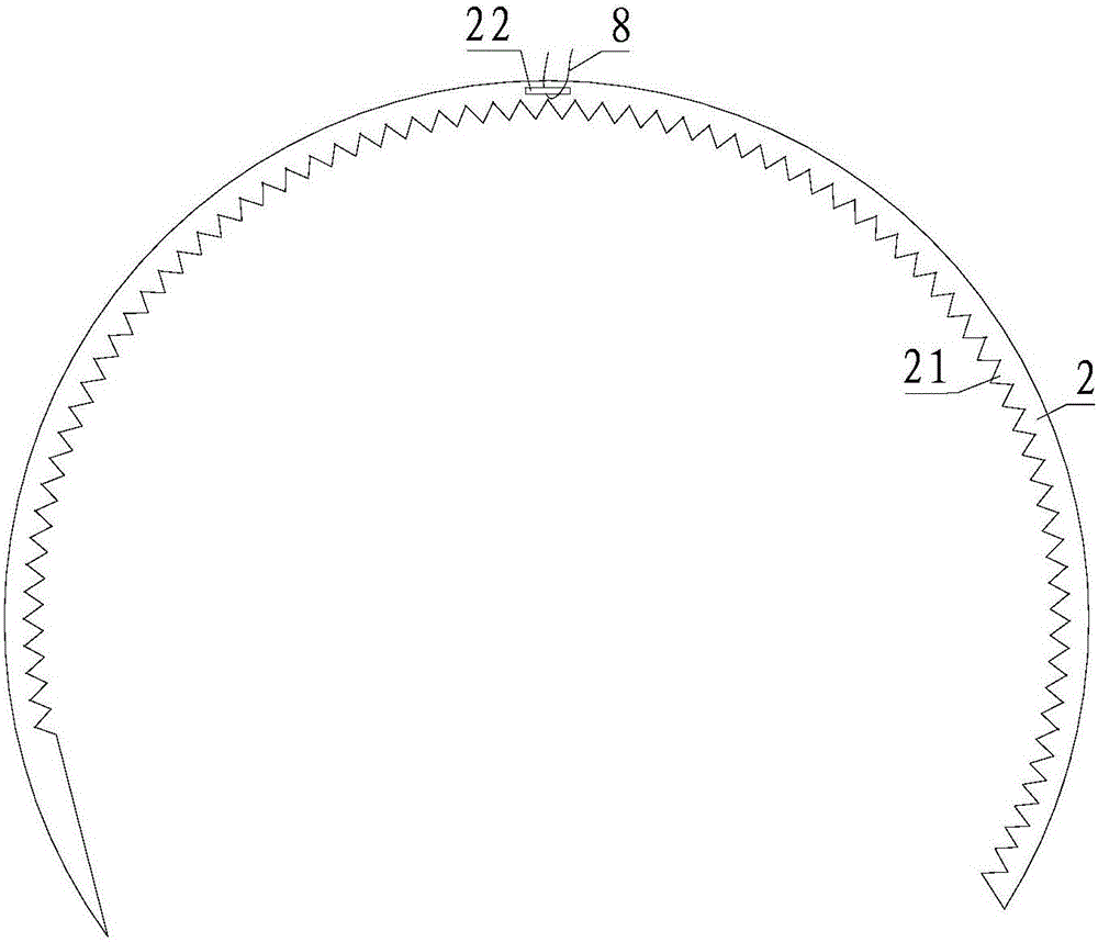
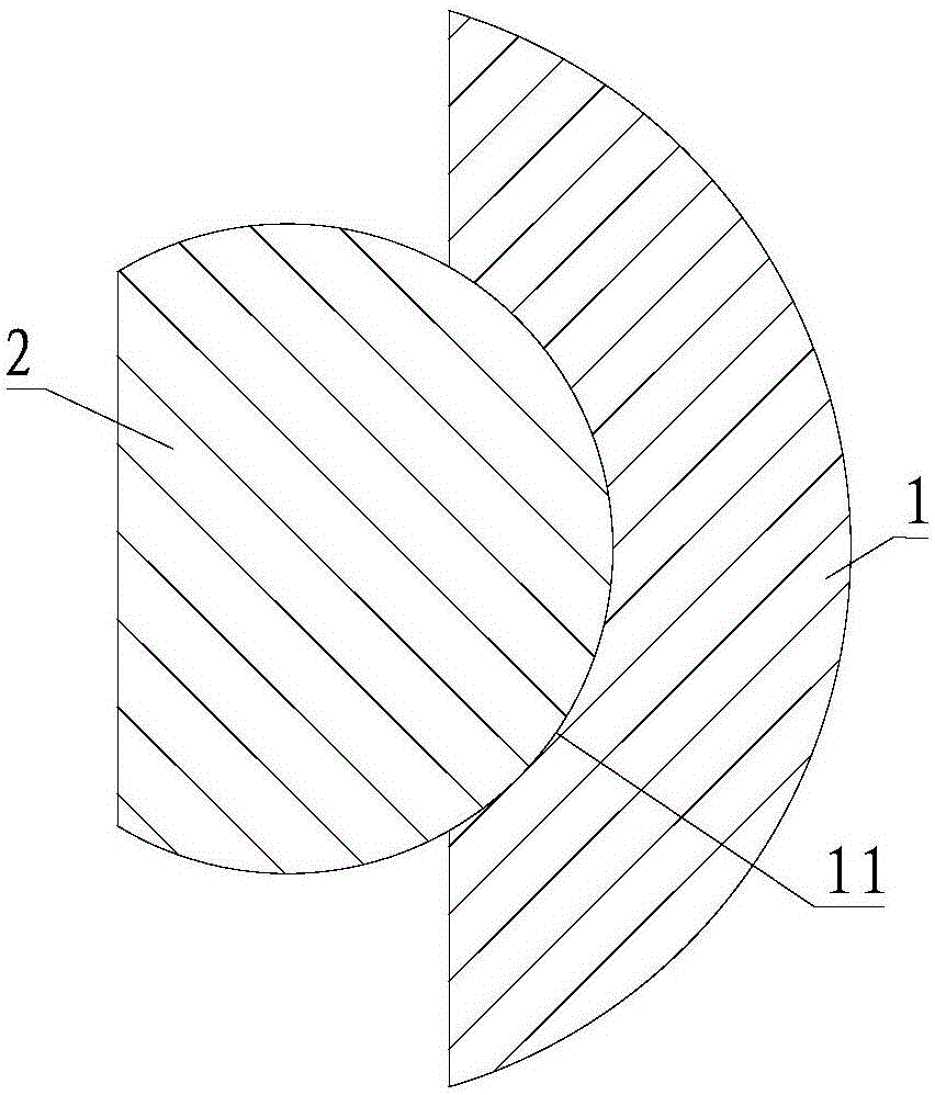
[0053] An endoscopic stapler, comprising an outer sheath 1, a suturing needle 2, a first drive assembly 3, a second drive assembly 4 and a steel wire 5, the projection shapes of the outer sheath 1 and the suture needle 2 are both C-shaped, the The inner surface of the outer sheath 1 is provided with an arc groove 11 adapted to the diameter of the suture needle 2, and the arc groove 11 is in contact with the outer surface of the suture needle 2, and the inner surface of the suture needle 2 Internal teeth 21 are provided, the first drive assembly 3 and the second drive assembly 4 are fixed on the outer sheath 1, and the ends of the motion transmission are all engaged with the internal teeth 21 on the suturing needle 2, the steel wire The end of 5 is Y-shaped, is divided into two branches, and is connected with the first drive assembly 3 and the second drive assembly 4 respectively, as the...

Embodiment 2

[0054] Please refer to figure 1 , the second embodiment of the present invention is:

[0055] An endoscopic stapler, on the basis of Embodiment 1, the first drive assembly 3 includes a first gear 31, a first ratchet 32, a first swing lever 33, a first active pawl 34, a first non-return The pawl 35 and the first spring 36, the second drive assembly 4 includes a second gear 41, a second ratchet 42, a second swing sleeve, a second active pawl 44, a second non-return pawl 45 and a second spring 46. ​​The first gear 31 and the second gear 41 are respectively located at the two ports of the outer sheath 1, and are both meshed with the internal teeth 21 of the suture needle 2, and the first ratchet 32 ​​and the second ratchet A gear 31 is set in coaxial linkage, the end of the steel wire 5 is branched to have two ends, one end of the first swing lever 33 is connected to one end of the end of the steel wire 5, and the other end is connected to the end of the first ratchet 32. One en...

Embodiment 3

[0056] Please refer to Figure 5 to Figure 8 , Embodiment three of the present invention is:

[0057] An endoscopic stapler, on the basis of Embodiment 1 or 2, further includes a sleeve 6 and a metal hose 73, the outer diameter of the sleeve 6 is adapted to the diameter of the external endoscope 9, so The sleeve 6 is installed at the end of the endoscope 9, the outer sheath 1 is installed in the inner hole of the sleeve 6, and the outer sheath 1 is arranged parallel to a longitudinal section of the sleeve 6, The opening of the outer sheath 1 faces the outside of the sleeve 6, and the sleeve 6 is provided with a through hole 61 on a side perpendicular to the longitudinal section of the outer sheath 1, and the steel wire 5 passes through the through hole 61. In the hole 61 , the steel wire 5 passes through the metal hose 73 , and the metal hose 73 is arranged side by side with the external endoscope 9 . The sleeve 6 is provided with a groove 62 on the surface opposite to the t...


PUM


Abstract

The invention provides an endoscope suture instrument comprising an sheath, a suture needle, a first driving component, a second driving component and a steel wire, wherein the projections of both the sheath and the suture needle are C-shaped, the sheath is provided with an arc-shaped groove fit with the diameter of the suture needle on the inner side, the arc-shaped groove is in contact with the outer side of the suture needle, inner teeth are arranged on the inner side of the suture needle, the first driving component and the second driving component are fixed on the sheath and movement transmission tail end of each of the first and second driving components is engaged with the inner teeth on the suture needle, and the tail end of the steel wire is Y-shaped and divided into two branches respectively connected with the first driving component and the second driving component to serve as power elements of the first driving component and the second driving component. The first driving component and the second driving component drive the suture needle to rotate to one direction in relays; the sheath plays a role in guiding the movement of the suture needle; the steel wire moves to and fro to driving the suture needle to rotate; the endoscope suture instrument is reasonable in structure, and the suture needle is good in continuous suture operability.

Description

technical field [0001] The invention relates to the technical field of medical instruments, in particular to an endoscopic stapler. Background technique [0002] The endoscopic stapler is used for suturing the internal operation of the gastrointestinal tract. When in use, the endoscopic stapler enters the gastrointestinal tract of the human body through the endoscope. The Chinese invention patent with the publication number CN105919636A discloses a rotatable stapler-type endoscopic suturing device, which includes a suture needle and a clip. The clip includes an open annular sleeve through which the suture needle can be put into the sleeve. Inside the barrel, the annular sleeve is provided with a fixed base and a movable suture needle baffle for opening and closing the opening. The baffle is connected to the base by a spring, and a pull cord is provided to drive the baffle to move. When the stapler is suturing the wound or tissue, turn the skin on both sides of the wound up ...

Claims


Application Information

Patent Timeline
Patent Type & Authority Applications(China)
IPC IPC(8): A61B17/04
CPCA61B17/0469
Inventor 李达周王雯李达龙
Owner 中国人民解放军联勤保障部队第九00医院
Why Eureka
Try Eureka