Automatic detection system for pulmonary nodule in chest CT (Computed Tomography) image

A CT imaging and automatic detection technology, applied in the direction of radiological diagnosis instruments, applications, image enhancement, etc., can solve problems such as the inability to meet the needs of doctors to judge pulmonary nodules, achieve broad market application prospects, and improve diagnostic accuracy Effect

Active Publication Date: 2017-05-31
杭州健培科技有限公司
View PDF3 Cites 172 Cited by
  • Summary
  • Abstract
  • Description
  • Claims
  • Application Information

AI Technical Summary

Problems solved by technology

These algorithms can no longer meet the needs of

Method used


Image

  • Automatic detection system for pulmonary nodule in chest CT (Computed Tomography) image
  • Automatic detection system for pulmonary nodule in chest CT (Computed Tomography) image
  • Automatic detection system for pulmonary nodule in chest CT (Computed Tomography) image

Examples


Embodiment Construction

[0030] The inventive concept of the present invention is to provide an "end-to-end" automatic pulmonary nodule detection solution.

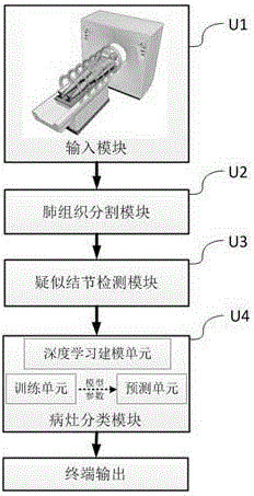
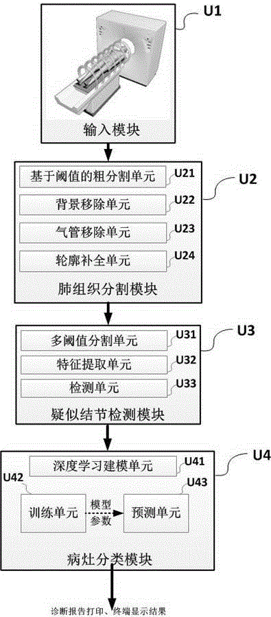
[0031] The present invention will be further described below in conjunction with the accompanying drawings and embodiments. In the following description, numerous specific details are set forth in order to provide a thorough understanding of the present invention. However, the invention can be carried out in many other ways than those described herein and thus the invention is not limited to the specific implementations disclosed below. figure 1 It is a structural schematic diagram of an automatic detection system for pulmonary nodules used in chest CT images according to the present invention.

[0032] Including: (1) Input module U1, used to acquire CT images, take lung CT image data through CT equipment, and input them to the pulmonary nodule detection system.

[0033] (2) The lung tissue segmentation module U2, configured to segment the lun...


PUM


Abstract

The invention relates to an automatic detection system for a pulmonary nodule in a chest CT (Computed Tomography) image. The system provides improvements for the problems of large calculated amount of computer aided software, inaccurate prediction and few prediction varieties. The improvement of the invention comprises the steps of acquiring a CT image; segmenting a pulmonary tissue; detecting a suspected nodular lesion area in the pulmonary tissue; classifying nidi based on a nidus classification model of deep learning; and outputting an image mark and a diagnosis report. The system has high nodule detection rate and relatively low false positive rate, acquires an accurate locating, quantitative and qualitative result of a nodular lesion and a prediction probability thereof. The end-to-end (from a CT machine end to a doctor end) nodular lesion screening is truly realized, the accuracy and operability demands of doctors are satisfied and the system has wide mar5ket application prospects.

Description

technical field [0001] The invention belongs to the technical field of computer-aided diagnosis of medical images, and in particular relates to an automatic detection system for pulmonary nodules used in chest CT images. Background technique [0002] The application of CT medical imaging can assist doctors in diagnosing whether a patient has lung cancer. However, the popularity of this application and the increase in the number of patients have increased the daily burden of reading images for hospital radiologists. At present, many computer-aided diagnosis researchers have invented a variety of computer-aided detection systems for pulmonary nodules in order to reduce the amount of film reading for doctors. Most of the system algorithms first use methods such as threshold segmentation, region growth, and edge detection to obtain CT images. The approximate area of ​​the middle lung parenchyma is then judged by performing true and false positives of lung nodules. These algori...

Claims


Application Information

Patent Timeline
IPC IPC(8): G06T7/00G06T7/11G06T7/136A61B6/00
CPCA61B6/5217G06T7/0012G06T2207/10081G06T2207/20081G06T2207/30061
Inventor 何林阳程国华严超孔海洋陈波季红丽
Owner 杭州健培科技有限公司
Why Eureka
Try Eureka