A 3D-printed in vitro auxiliary positioning device for pulmonary masses and its preparation method

A 3D printing and assisted positioning technology, which is applied in the direction of stereotaxic surgical instruments, etc., can solve the problems of long time-consuming preoperative positioning methods, untouchable touch by hand touch, and inability to completely remove lung masses, so as to avoid inaccessible nodules, The effect of shortening operation time and reducing radiation damage

Active Publication Date: 2018-11-23
SHANGHAI PULMONARY HOSPITAL
View PDF4 Cites 0 Cited by
  • Summary
  • Abstract
  • Description
  • Claims
  • Application Information

AI Technical Summary

Problems solved by technology

[0003] However, there are the following technical problems in the prior art: a. The mass cannot be touched by the hand touch method in the operation: it is theoretically the most direct and accurate method to confirm the position of the mass by touching in the operation, but in many cases the operation is caused by the lung The texture of the mass is tough, the mass is too small, the position of the mass is deep, and the doctor is inexperienced, etc., which will lead to the inability to touch or the inaccurate touch of the hand, so that the lung mass cannot be completely removed; b. The influence of radiation radiation: surgery The puncture positioning guided by the hook-wire positioning needle or micro-coil micro-coil in the anterior CT scan requires multiple CT scans in a short period of time to determine the location and cause radiation impact on the patient; c. Staffing and cost: CT guides puncture positioning through hook-wire positioning needle or micro-coil microcoil, requiring at least two experienced medical staff to operate
[0004] The existing preoperative positioning method is time-consuming, requiring the patient to lie on the side of the CT bed in a fixed position for a long time, and undergo repeated adjustments and multiple punctures of the puncture needle, which is very painful
For patients who are unconscious or seriously ill and cannot cooperate, the existing technology is completely unfeasible

Method used


Image

  • A 3D-printed in vitro auxiliary positioning device for pulmonary masses and its preparation method
  • A 3D-printed in vitro auxiliary positioning device for pulmonary masses and its preparation method

Examples


preparation example Construction

[0042]This embodiment also provides a preparation method of the 3D printed lung mass in vitro auxiliary positioning device according to claim 1, comprising the following steps:

[0043] (1) According to the pre-processed positioning slice image with the lung mass, use modeling software to reconstruct the model of the patient's chest cavity and lungs with mass, and determine the projection of the lung mass on the body surface on the chest cavity and lung models Location;

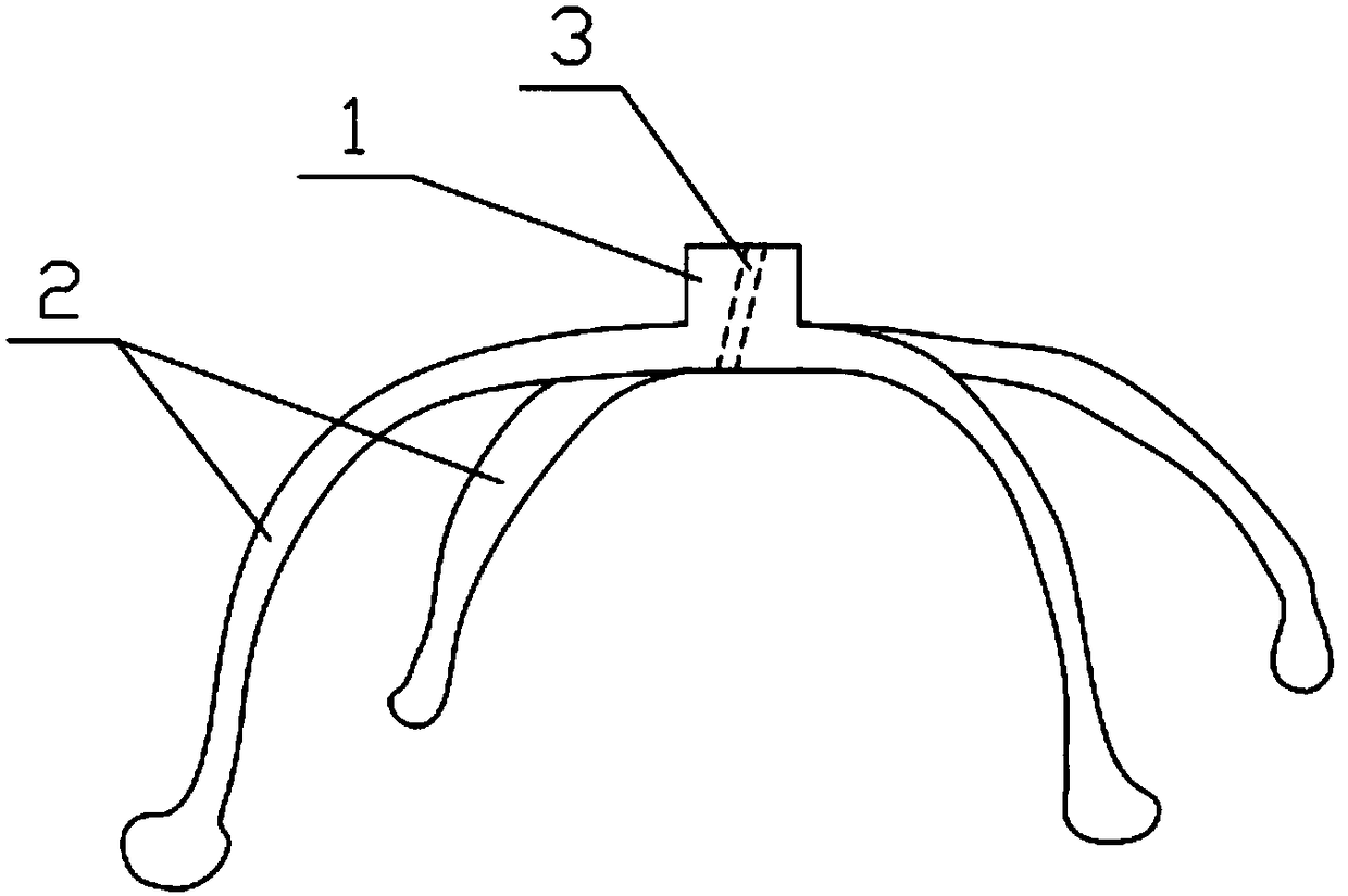
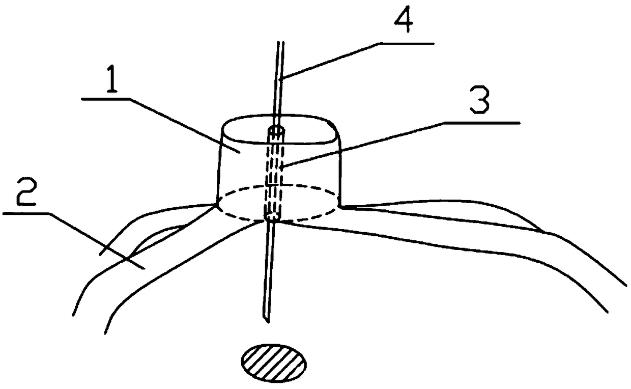
[0044] (2) According to the pre-calculated needle insertion angle and depth, use the modeling software again to build a fitted umbrella structure model including the legs 2 and the central positioning module 1 on the above-mentioned chest cavity and lung models;

[0045] (3) Import the umbrella structure model into the printer and perform 3D printing to obtain an auxiliary positioning device in vitro.

[0046] Wherein, the step (1) positioning slice image includes the position, size, depth and number of the ...


PUM


Abstract

The invention discloses a 3D-printed in-vitro assistant positioning device for a lung mass and a preparation method for the same. The positioning device is fabricated by 3D printing and comprises a central positioning module and supporting legs, wherein a needle inlet duct is formed in the central positioning module and used to restrain marking of a puncture needle aiming at the lung mass and stabilize a needle inlet direction of the puncture needle; and the supporting legs are bent, front ends of the supporting legs are firmly connected to the central positioning module, tail ends of the supporting legs are bent downwards and contacted with a human body, the multiple supporting legs are disposed and can support the central positioning module upwards to form an umbrella-shaped structure with the supporting legs as an umbrella frame, with the central positioning module as the top part and with the puncture needle as an umbrella rod. The invention provides the personalized lung mass assistant positioning device outside a chest wall, wherein the device is fabricated through three-dimensional reconstruction technologies and 3D printing technologies. The device has the advantages that positioning is accurate; problems in that nodules cannot be touched through a hand-touch method can be avoided; exposure and radiation damage brought by CT of a patient are reduced; operation duration is shortened; and medical resources are utilized effectively.

Description

technical field [0001] The invention relates to a positioning device for a pulmonary mass in laparoscopic surgery, in particular to a 3D printed external auxiliary positioning device for a pulmonary mass and a preparation method thereof. Background technique [0002] Various pulmonary masses, including pulmonary ground-glass nodules (GGO), are one of the surgical indications for thoracoscopic lung surgery. In the case of a tumor that is too small and deep, the location of the tumor cannot be directly seen during the operation, which affects the judgment of the resection site. At present, to solve this clinical problem, there are mainly three methods to determine the location of the lung mass: 1. Intraoperative exploration and positioning: During the operation, use hand touch to determine the location of the pulmonary nodule according to the approximate range of the pulmonary nodule on chest CT; 2. .Pulmonary nodules were located through the chest wall hook-wire positioning ...

Claims


Application Information

Patent Timeline
Patent Type & Authority Patents(China)
IPC IPC(8): A61B90/10
Inventor 李泽遥李牧王轶灵张旭峰王龙范子文顾亚伟陈昶
Owner SHANGHAI PULMONARY HOSPITAL
Why Eureka
Try Eureka