An autonomous fundus photography imaging system and method

A fundus photography and imaging system technology, applied in ophthalmoscopes, eye testing equipment, medical science, etc., can solve the problems of poor stability and repeatability, and achieve system energy saving, high precision, flexible and fast alignment of the visual axis Effect

Active Publication Date: 2018-11-02
SHANGHAI UNIV OF MEDICINE & HEALTH SCI
View PDF5 Cites 0 Cited by
  • Summary
  • Abstract
  • Description
  • Claims
  • Application Information

AI Technical Summary

Problems solved by technology

These methods all need to be equipped with additional personnel to manually operate the fundus camera system, and they all have problems such as poor stability and repeatability

Method used


Image

  • An autonomous fundus photography imaging system and method
  • An autonomous fundus photography imaging system and method
  • An autonomous fundus photography imaging system and method

Examples


Embodiment

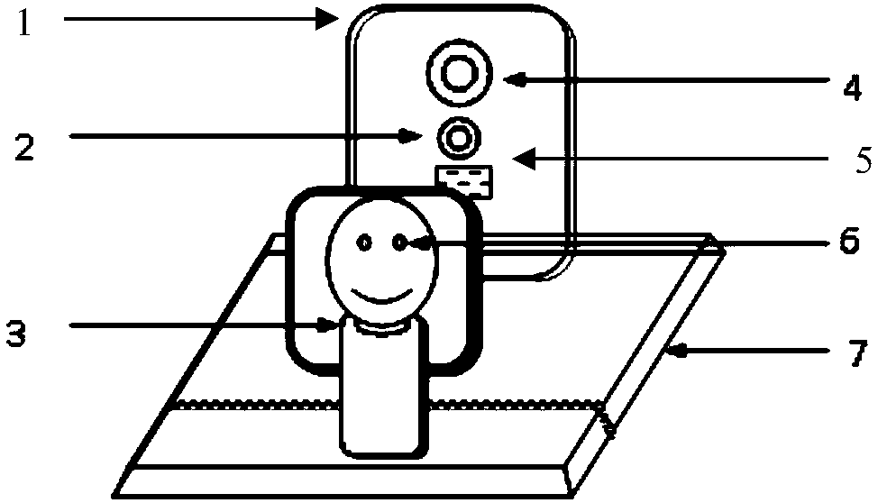
[0035] Such as figure 1 As shown, an autonomous fundus photography imaging system includes a fundus camera device 1, an auxiliary precise positioning device for pupils and a mandibular support device 3, and the fundus camera device 1 includes a fundus camera 4 fixed on a mobile working platform 7 and an optical positioning sensor 2. The eye pupil auxiliary precise positioning device includes a controller and a driving mechanism. The controller is connected to the driving mechanism, the fundus camera 4 and the optical positioning sensor 2 respectively, the driving mechanism is connected to the mobile working platform 7, and the mandibular support device 3 is connected to the fundus camera device 1. Relatively set, when working, the optical positioning sensor 2 takes pictures of the eyes of the measured object and the mandibular support device 3, and the controller calculates the distance between the mandibular support device 3 and the fundus camera 4 and the distance between the...


PUM


Abstract

The invention relates to an autonomic fundus photography imaging system and method. The autonomic fundus photography imaging system comprises a fundus photographing device, a pupil auxiliary accurate locating device and a lower jaw supporting system (3), the fundus photographing device comprises a fundus camera (4) and a optical locating sensor (2) which are fixed on a mobile operation platform (7), the pupil auxiliary accurate locating device comprises a controller and a driving mechanism, the controller is separately connected with the driving mechanism, the fundus camera (4) and the optical locating sensor (2), the driving mechanism is connected with the mobile operation platform (7), and the lower jaw supporting device (3) is arranged opposite to the fundus photographing device. Compared with the prior art, the autonomic fundus photography imaging system based on the existing fundus photographing method, is additionally provided with the pupil auxiliary accurate locating device and the optical locating sensor, achieves double-eye searching and optical axis alignment, conducts autonomic accurate fundus photographying, and has the advantages of high efficiency and accuracy.

Description

technical field [0001] The invention relates to a fundus imaging device, in particular to an autonomous fundus imaging system and method. Background technique [0002] Fundus photographic imaging inspection is usually performed by professional ophthalmologists using a precision optical fundus camera to observe and record the physiological structure of the fundus through manual assisted positioning and focusing. Fundus photography can directly observe the capillaries and optic nerve structures distributed on the fundus retina, so fundus photography inspection is of great significance for early diagnosis and treatment follow-up of fundus diseases, diabetes and hypertension. [0003] Since the German Zeiss company successfully developed the fundus imaging technology in 1925, the fundus imaging methods have basically followed the coaxial illumination optical system. During the shooting process, professionals are required to assist the subject, manually locate and focus the fund...

Claims


Application Information

Patent Timeline
Patent Type & Authority Patents(China)
IPC IPC(8): A61B3/12A61B3/14
CPCA61B3/0008A61B3/0016A61B3/0075A61B3/0083A61B3/12A61B3/14A61B3/152A61B3/154
Inventor 黄钢孙九爱何培忠姚旭峰刘红
Owner SHANGHAI UNIV OF MEDICINE & HEALTH SCI
Why Eureka
Try Eureka