Deep learning-based ultrasound contrast tumor automatic identification and detection method

A contrast-enhanced ultrasound and automatic recognition technology, applied in the field of image recognition, can solve the problems of low segmentation accuracy, inability to achieve high accuracy in classifier recognition, and inability to achieve prediction, and achieve the effect of good segmentation accuracy.

Inactive Publication Date: 2017-07-28
CHONGQING UNIV
View PDF0 Cites 21 Cited by
  • Summary
  • Abstract
  • Description
  • Claims
  • Application Information

AI Technical Summary

Problems solved by technology

However, due to the diversity of lesion shapes, it is impossible to achieve effective prediction when dealing with unknown types of lesions.
Among them, the recognition accuracy rate for liver cancer tissues with different tissue textures is only 65.2%, and the recognition rate for completely different types of liver cancer tissues is only 41.7%. Based on existing traditional classifiers such as decision trees, multi-classification logistic regression, etc., Segmentation accuracy is low, with high false positive rate and false negative rate; existing classifiers cannot achieve high accuracy in recognition
[0003] Ultrasound-enhanced tumor detection is one of the most sensitive and accurate detection methods in tumor detection. However, the existing ultrasound-enhanced tumor diagnosis is based on the professional knowledge of doctors and has high requirements for doctors' professional level. Therefore, there are only a few high-quality methods. Level hospitals can carry out ultrasound-enhanced tumor detection

Method used


Image

  • Deep learning-based ultrasound contrast tumor automatic identification and detection method
  • Deep learning-based ultrasound contrast tumor automatic identification and detection method
  • Deep learning-based ultrasound contrast tumor automatic identification and detection method

Examples


Embodiment 1

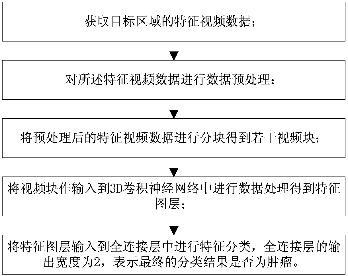
[0029] The deep learning-based method for automatic identification and detection of tumors in contrast-enhanced ultrasound provided in this embodiment includes the following steps:

[0030] Acquiring characteristic video data of the target area; the characteristic video is a video that is stable for a period of time after the microbubbles are injected into the tissue area completely filled with the microbubbles;

[0031] Perform data preprocessing on the feature video data:

[0032] Preprocessing: The original video is video data with a frame rate of 100 frames per second and a duration of 200 seconds. A video has a size of 2GB. Direct processing of such a large amount of data will result in too long training and testing time. The data of 1 key frame per second injected into the filling tissue for 10 seconds is used as the video data obtained by preliminary preprocessing. Among them, it is necessary to ensure that the frames intercepted between different samples have the same...

Embodiment 2

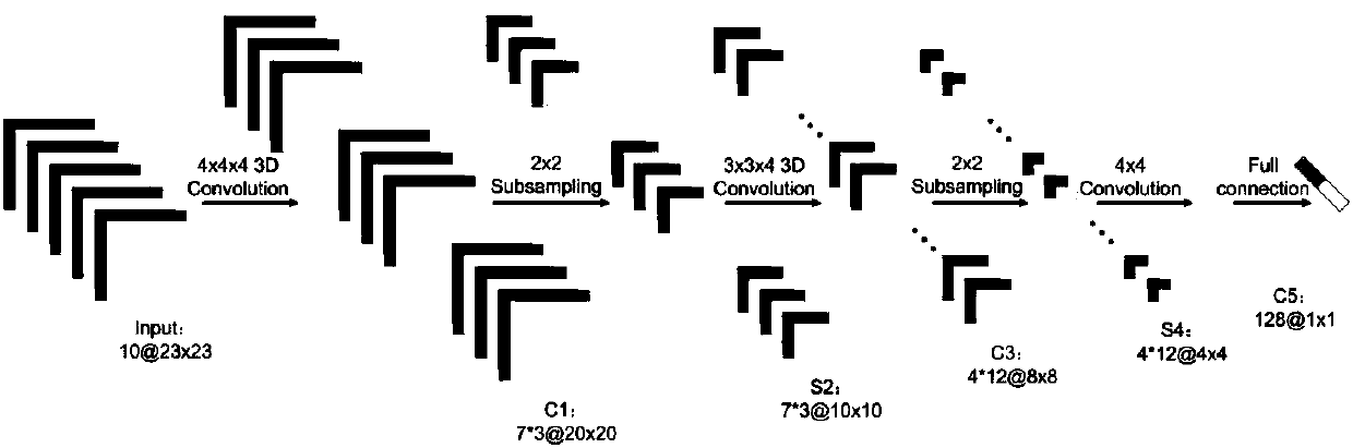
[0044] The deep learning-based automatic recognition and detection method for tumor contrast-enhanced ultrasound provided in this embodiment uses a 3D convolutional neural network to effectively extract the time-space effective blood flow characteristics within and between frames of contrast-enhanced ultrasound video data, and based on the tumor The difference between tissue and normal tissue can realize the detection of tumor tissue. This method takes into account both the spatial information around the target area and the time-varying information, which improves the segmentation accuracy; takes into account the surrounding spatial information and continuous time information; effectively integrates the nano-microbubbles in the tissue (or tumor) Flow process information is better used for tumor region segmentation. To achieve a higher segmentation accuracy, the specific steps are as follows:

[0045] Data preprocessing: intercept the video, and intercept the feature video dat...


PUM


Abstract

The present invention discloses a deep learning-based ultrasound contrast tumor automatic identification and detection method. The method comprises the following steps of obtaining the characteristic video data of a target area; pre-processing the characteristic video data; partitioning the pre-processed characteristic video data to obtain a plurality of video blocks; inputting the video blocks in a 3D convolutional neural network to process the data and obtain a characteristic layer; inputting the characteristic layer in a full-connection layer to classify the characteristics, and finally determining whether the classification result is a tumor. By utilizing the 3D convolutional neural network, the method provided by the present invention can effectively extract the time-space effective blood flow characteristics in and between the ultrasound contrast video data frames, detects the tumor tissues based on the difference of the tumor tissues and the normal tissues and by combining the time-space characteristics of the video data and using a 3D CNN algorithm to segment the tumors, and has a better segmentation accuracy than a conventional machine learning algorithm.

Description

technical field [0001] The invention relates to the technical field of image recognition, in particular to a method for automatic recognition and detection of tumors in contrast-enhanced ultrasound based on deep learning. Background technique [0002] At present, there are few studies on computer-aided diagnosis methods for tumor diagnosis by contrast-enhanced ultrasound. By searching related keywords, it is found that there are few studies on this method in recent years. The main research idea is to use B-ultrasound images and known liver lesions. Images are used as data, and human neural networks and decision trees are used for classification, achieving a certain degree of accuracy. However, due to the diversity of lesion shapes, effective prediction cannot be achieved when dealing with unknown types of lesions. Among them, the recognition accuracy rate for liver cancer tissues with different tissue textures is only 65.2%, and the recognition rate for completely different...

Claims


Application Information

Patent Timeline
Patent Type & Authority Applications(China)
IPC IPC(8): G06K9/62G06N3/04G06N3/08
CPCG06N3/084G06N3/045G06F18/241
Inventor 周喜川杨帆冯玉洁谭跃李胜力李坤平赵昕刘念徐埌
Owner CHONGQING UNIV
Why Eureka
Try Eureka