Microfluidic blood coagulation detection device and method

A detection device and detection method technology, applied in the field of microfluidics, can solve problems such as large amount of reagents, and achieve the effects of shortening detection time, speeding up the process of reaction, and process efficiency

Active Publication Date: 2017-08-18
ZHEJIANG PUSHKANG BIOTECHNOLOGY CO LTD
View PDF8 Cites 14 Cited by
  • Summary
  • Abstract
  • Description
  • Claims
  • Application Information

AI Technical Summary

Problems solved by technology

[0008]The amount of reagents used in the existing tubular blood coagulation is relatively large. For projects with high reagent costs, reducing the amount of reagents used can significantly reduce the detection cost of users, especially For primary medical institutions, they need miniaturized and simplified inspection equipment

Method used


Image

  • Microfluidic blood coagulation detection device and method
  • Microfluidic blood coagulation detection device and method
  • Microfluidic blood coagulation detection device and method

Examples


Embodiment 1

[0030] The microfluidic hemagglutination detection device according to the embodiment of the present invention, the microfluidic hemagglutination detection device includes:

[0031] An optical detection unit, the optical detection unit is a prior art in the field, and the specific structure and detection methods will not be repeated here;

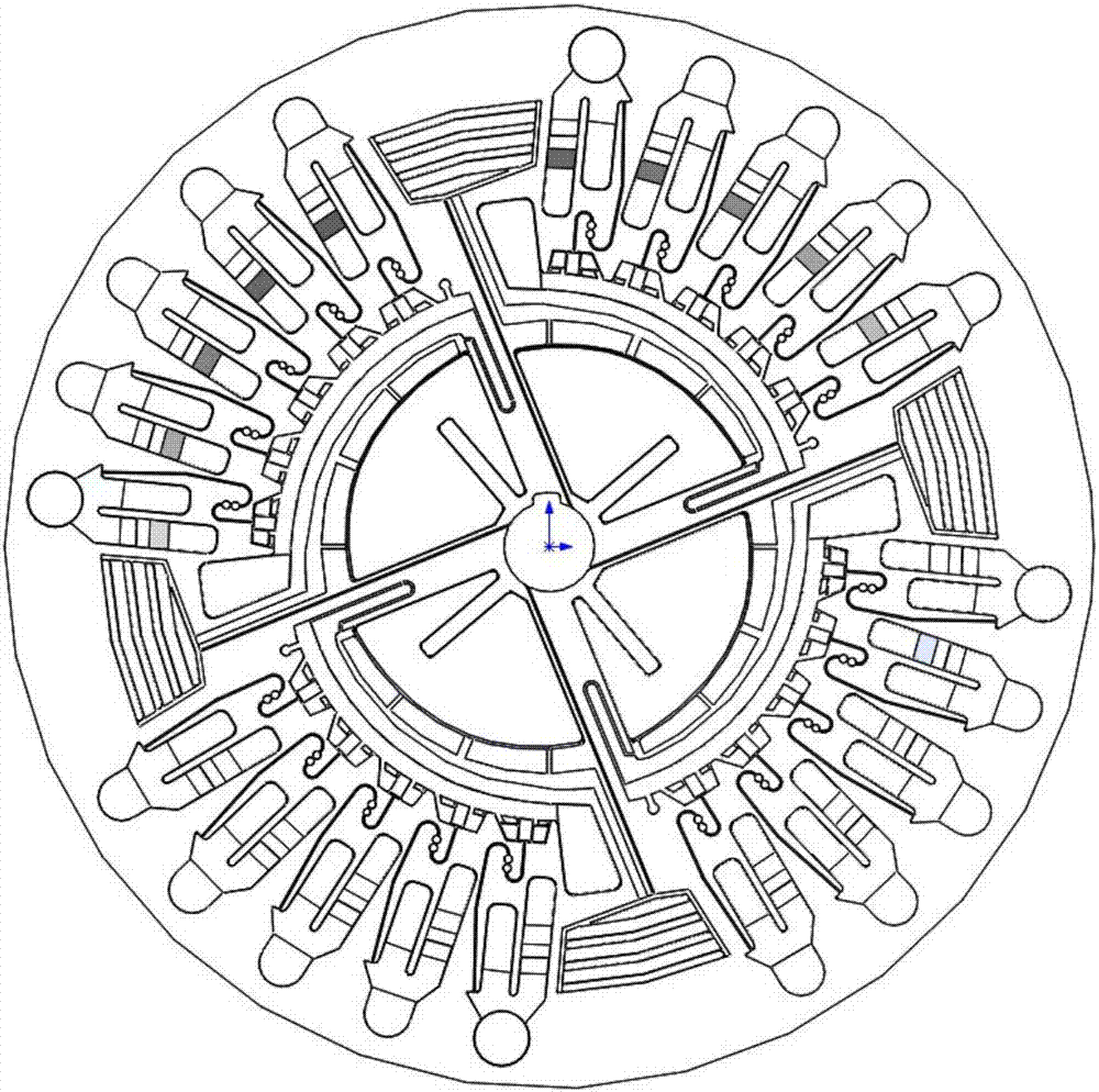
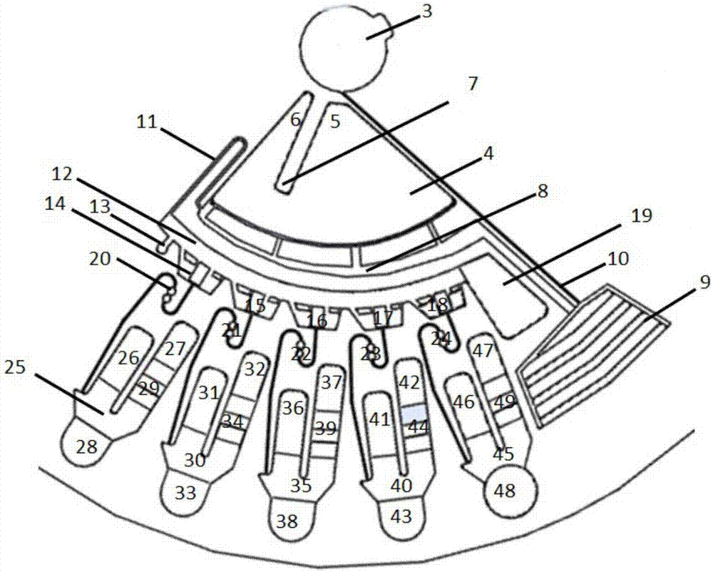
[0032] figure 1 Schematically shows the structural diagram of the disk of the embodiment of the present invention, such as figure 1 As shown, the disk includes a plurality of microchannel units, such as 4, such as figure 2 As shown, each microfluidic unit includes:

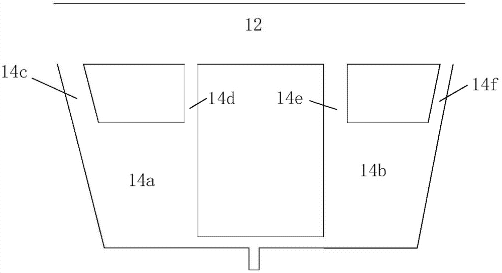
[0033] Injection groove 4, described injection groove has sample loading hole 5, exhaust hole 6 and dividing plate 7, and described sampling hole and emptying hole are separated by described dividing plate;

[0034] The first channel 8, the first channel is distributed along the circumferential direction of the disc, and the width in the radial direction of the disc gradual...

Embodiment 2

[0056] The microfluidic hemagglutination detection method of the embodiment of the present invention is different from Embodiment 1 in that:

[0057] Step (A5) is specifically:

[0058] The reagent is added into the sample loading area of ​​the sample loading tank, the disk rotates, the reagent enters the reaction area through the communication area, and at the same time, the plasma in the quantitative tank breaks through the third channel and enters the reaction area of ​​the sample loading tank.

Embodiment 3

[0060] An application example of the microfluidic hemagglutination detection device and working method according to Embodiment 1 of the present invention in the detection of whole blood samples.


PUM


Abstract

The invention provides a microfluidic blood coagulation detection device and method. The microfluidic blood coagulation detection device includes a disc, the disc comprises micro-channel units, each micro-channel unit comprises: an injection groove, which is equipped with a sample adding hole, a vent hole and a partition plate, wherein the sample adding hole and the vent hole are separated by the partition plate; a first channel, which is distributed along the circumferential direction of the disc, has a gradually increasing width in the radial direction of the disc, and is located at the periphery of the injection groove that is communicated with a storage groove through the first channel; communication channels, which are used for communicating with the injection groove and the first channel and are disposed in the radial direction of the disc; the storage groove, which is further away from a rotation center of the disc compared with the first channel and is communicated with a widest part of the first channel. The device and the method provided by the invention have the advantages of high efficiency, stability and simplicity, etc., and can achieve direct injection of a whole blood sample.

Description

technical field [0001] The invention relates to microfluidics, in particular to a microfluidic hemagglutination detection device and method. Background technique [0002] At present, the popular automatic tube coagulation detection process is as follows: [0003] 1. Add plasma samples; [0004] 2. Add reagent 1; [0005] 3. Mix the sample and reagent 1; [0006] 4. Add reagent 2; [0007] 5. Mix and test. [0008] The amount of reagents used for the existing tubular coagulation is relatively large. For projects with high reagent costs, reducing the amount of reagents used can significantly reduce the testing cost of users, especially for grassroots medical institutions, who need miniaturized and simplified testing. equipment. Contents of the invention [0009] In order to solve the deficiencies in the above-mentioned prior art solutions, the present invention provides an efficient, stable and easy-to-operate microfluidic blood coagulation detection device. [0010] ...

Claims


Application Information

Patent Timeline
Patent Type & Authority Applications(China)
IPC IPC(8): B01F13/00B01F11/00B01L3/00G01N33/86
CPCG01N33/86B01L3/5027B01F31/10B01F33/30B01F2101/23
Inventor 刘志远陈坦金烨琦程林余波
Owner ZHEJIANG PUSHKANG BIOTECHNOLOGY CO LTD
Why Eureka
Try Eureka